





















 /SC-1600

 TOP
TOP


Optical Coherence Tomography / Fundus Camera
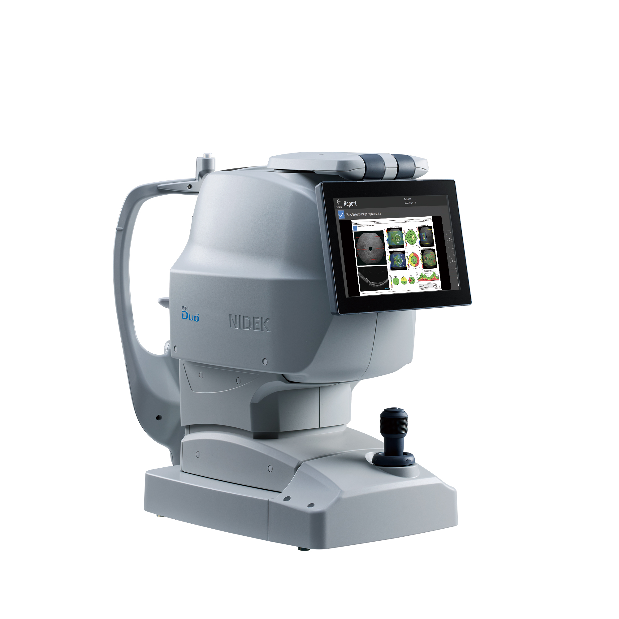
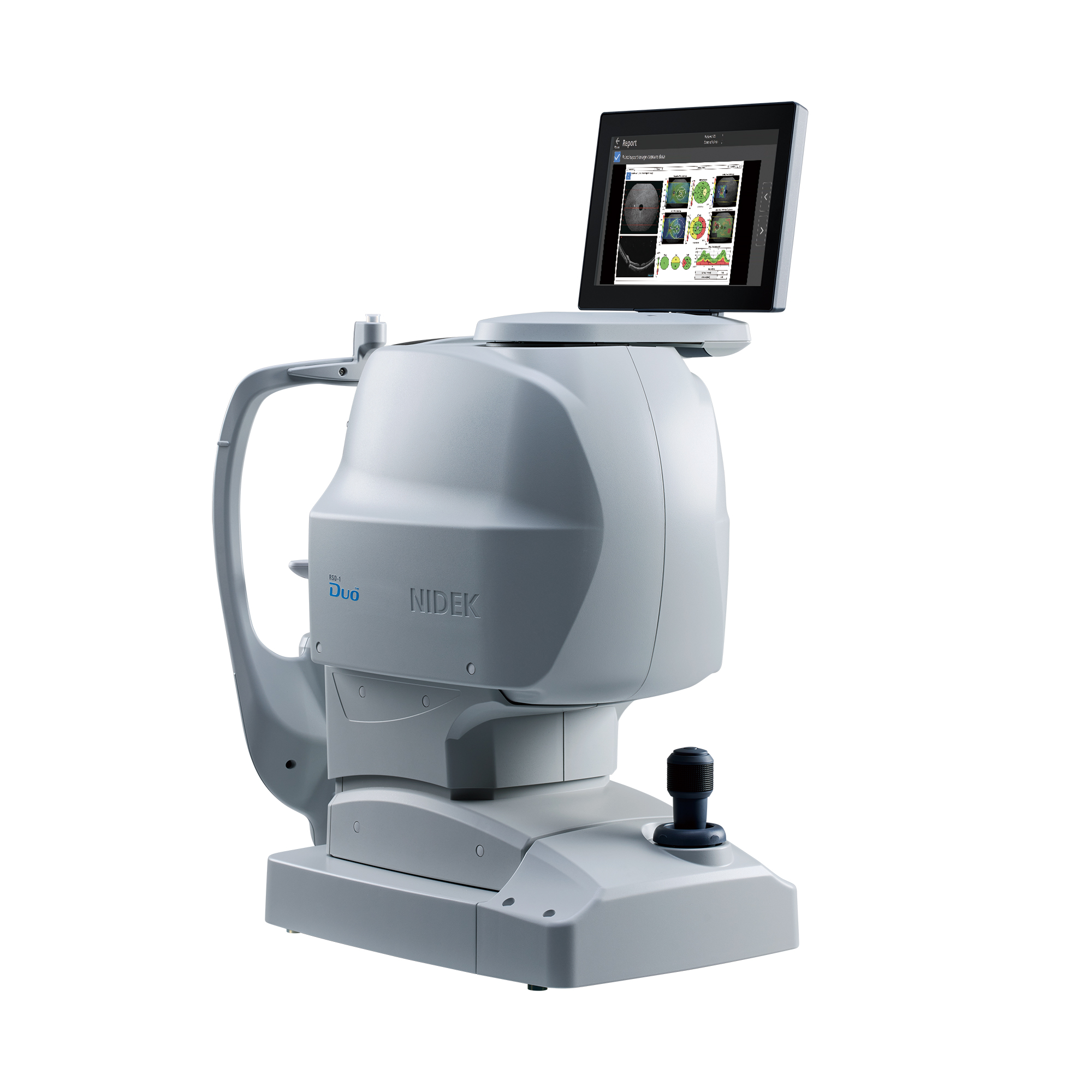
RSD-1 Duo™
Features
- Five auto functions achieving universal operation
- Wide and deep B-scan with denoising technique
- 15 x 10 mm retina map
- Axial length compensation analysis
- Enhanced OCT-Angiography* with advanced imaging algorithm
- Slit-scan fundus photography
- FAF**
- Flexible installation & data management
*Optional
**Available for the FAF model
Detailed Information
Five auto functions achieving universal operation
| The five auto functions allow anyone to simply capture images. The RSD-1 Duo continuously captures OCT and fundus images and switches to small pupil mode automatically when needed, enhancing clinical workflow. | 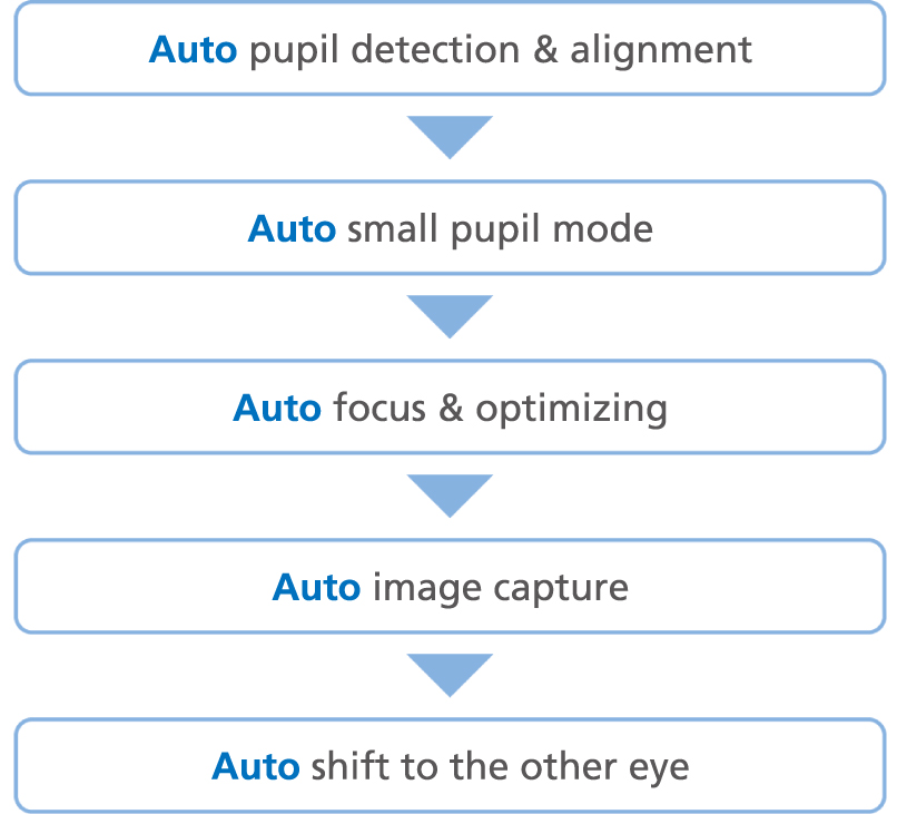 |
Wide and deep B-scan with denoising technique
Wide and deep B-scanWith the RSD-1 Duo, a single B-scan image clearly presents the area from the optic nerve head to the temporal vascular arcade, and the 3.0 mm depth B-scan imaging readily captures the oblate retinal shape of myopic eyes. |
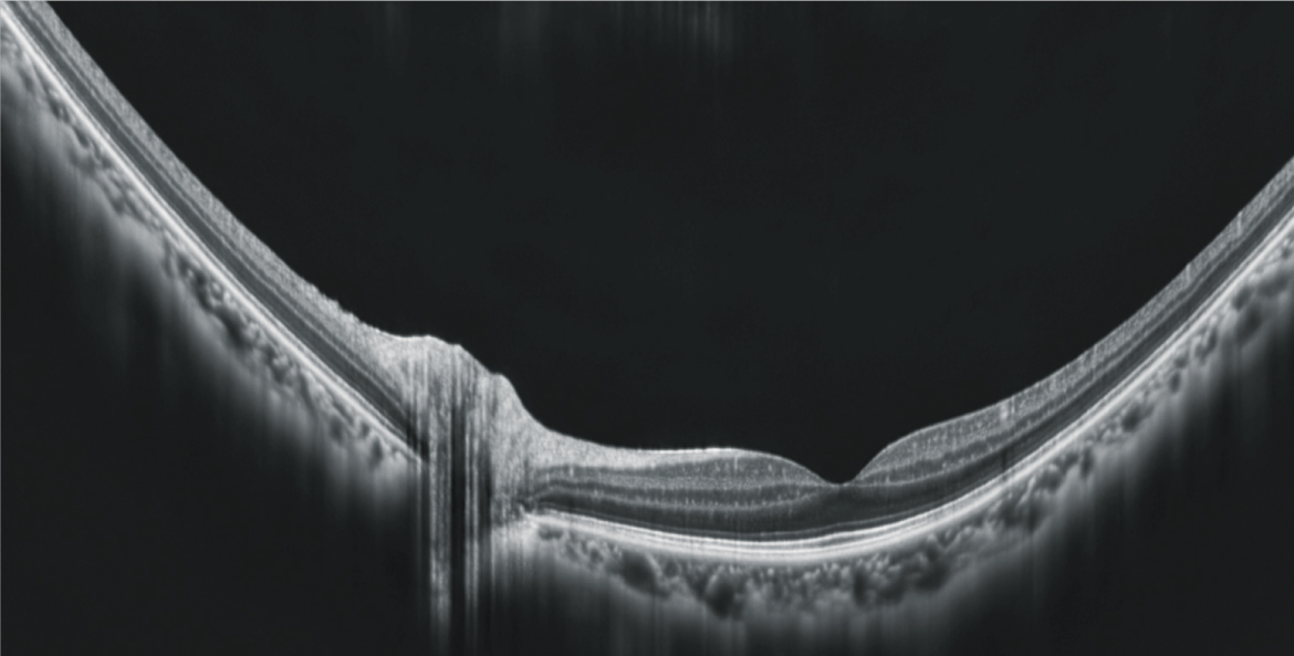
Image after B-scan denoise Macula line: 16.5 mm / Scan depth: 3.0 mm |
B-scan denoise delivers image-averaging quality from a single frameWith deep learning of a data set of images averaged from 120 images, this denoising technique provides high-definition images comparable to a multiple-image-averaging technique. When working without EyeLink, the RSD-1 Duo can denoise a selected single B-scan image. |
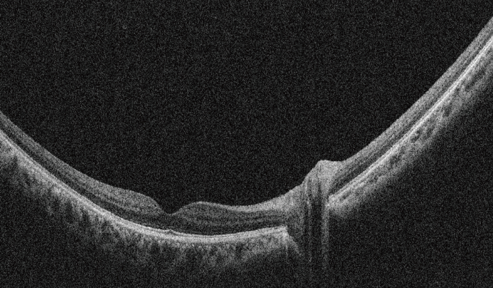 Single-frame B-scan image Single-frame B-scan image |
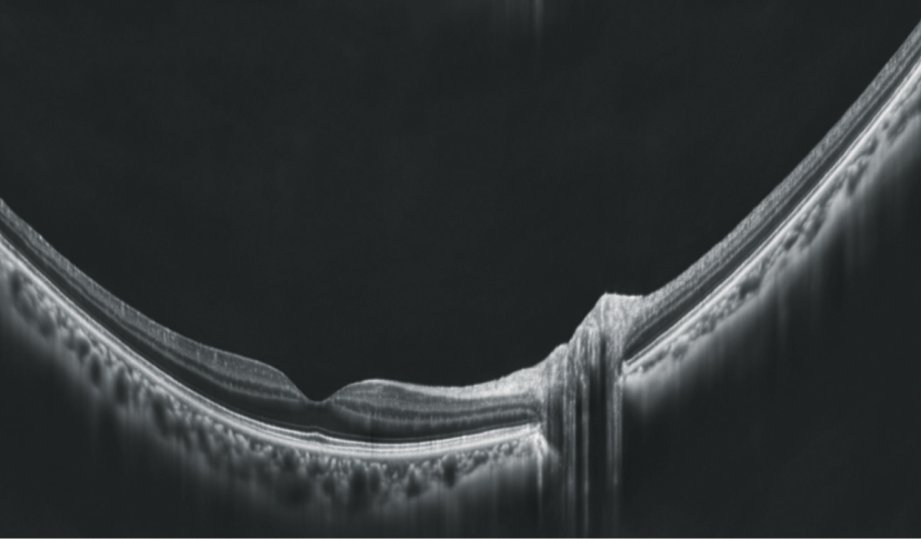
Image after B-scan denoise |
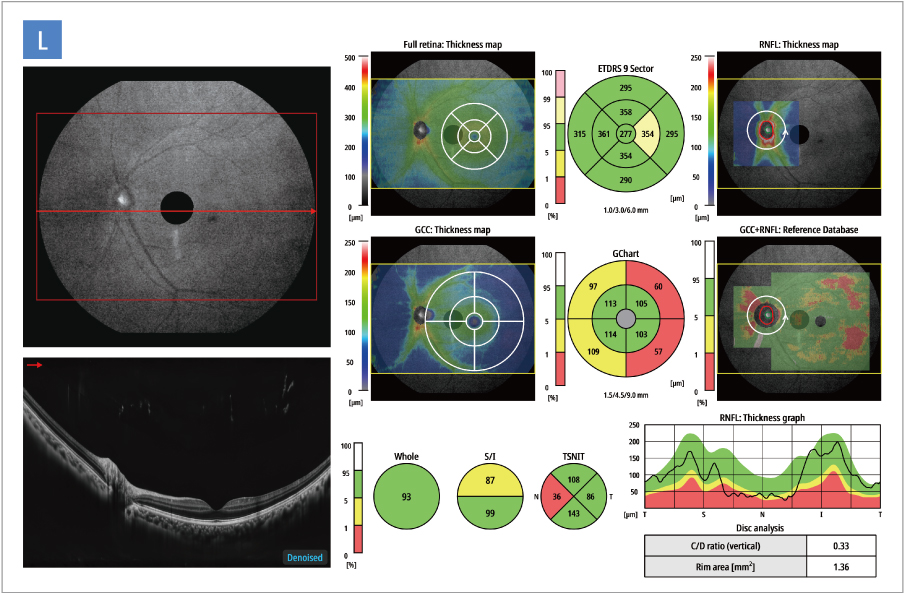
15 x 10 mm retina map
| The retina map captures both the disc and macula in a single shot. Reference database* analysis is performed for both the RNFL and GCC on a single OCT image, facilitating efficient diagnostic screening.
*Data was collected from a sample of Caucasian and Asian patients. |
 |
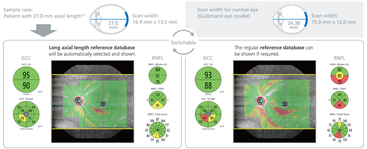
Axial length compensation analysis
| The long axial length reference database (LAL-RDB)*1 presents analysis with axial length compensation, facilitating for more accurate glaucoma assessment in patients with axial myopia. The RSD-1 Duo automatically switches to this database as required, by using the axial length*2 which is a parameter for scan width correction.
*1 Data was collected from a sample of Asian patients. *2 The value of the axial length is obtained based on the results of the OCT image capture and differs from the actual measured value of the axial length. |
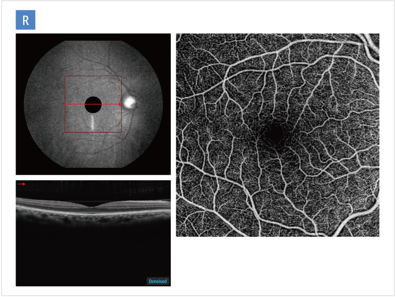
Enhanced OCT-Angiography with advanced imaging algorithm
| Complex OCT signal Difference Analysis Angiography 2 (CODAA 2), improves contrast, allowing for enhanced imaging. The high-definition OCT-Angiography (optional) enables greater clinical use of noninvasive imaging of retinal vascular diseases, reducing dependence on contrast dye tests.
Scan size – Macula map: 6 x 6 mm – Disc map: 6 x 6 mm |
 |
Slit-scan fundus photography
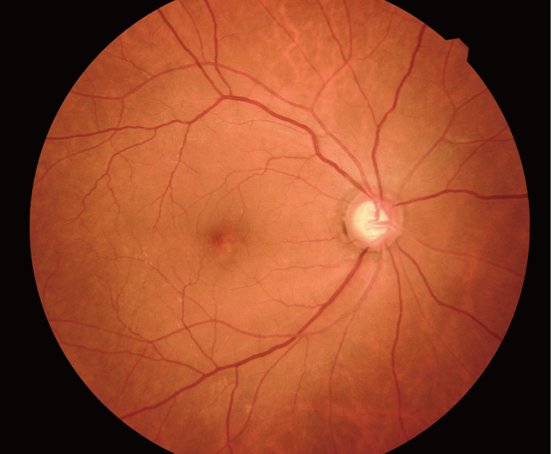
High-contrast imaging even for challenging casesThe advanced slit-scan technology delivers high-contrast, high-definition images with fewer artifacts and shadows. Despite room-light conditions and pupil constriction, it achieves a higher imaging success rate, including in cases previously considered difficult, such as cataracts or small pupils (min. ø2.0 mm).
Consistent wide imagingThe RSD-1 Duo captures up to 50° wide fundus images (converted to 74° at the center of the eye). Even in auto small pupil mode, the angle of view remains 50°, ensuring consistent performance for every patient. |
 |
PanView—Panorama imaging like never before
| Seamless auto-composite panorama imaging delivers stunning clarity, far surpassing the previous model. The RSD-1 Duo can complete panorama composition quickly on the device, reducing operator time and effort. With the ability to automatically combine up to seven shots, the RSD-1 Duo produces expansive panoramic fundus images of up to 98° (converted to 145° at the center of the eye). | 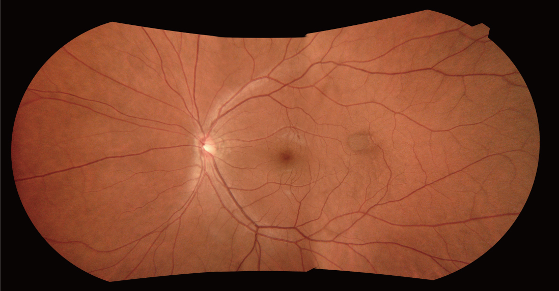 |
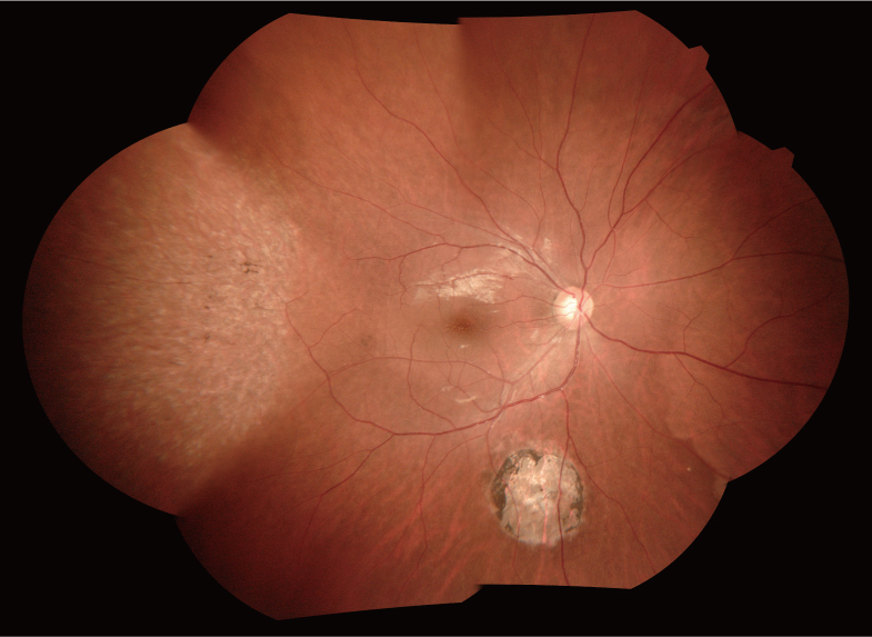 |
The angle of view measured at the center of the eye: The angle of view measured at the center of the eye is a reference value obtained by converting the field of view specified in ISO 10940:2009 into an angle subtended by the endpoints of the captured area at the center of a simplified eye model. Note that this conversion is based on a simplified approximation and therefore differs in definition from the field of view specified in ISO 10940:2009. The angle of view measured at the center of the eye differs from the actual field of view for image capture.
The field of view of the panorama image: The field of view of the panoramic composition is defined as the single-shot field of view, in accordance with ISO 10940:2009, and the maximum angle that accounts for gaze deviation induced by internal fixation guidance. The actual field of view of the panoramic composition may vary depending on patient fixation, imaging conditions, and image composition parameters.
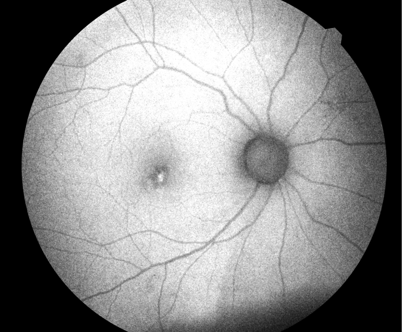
FAF
| The FAF* function is an advanced screening feature that enables non-invasive evaluation of the RPE without contrast dye.
*Available for the FAF model |
 |
Flexible installation & data management
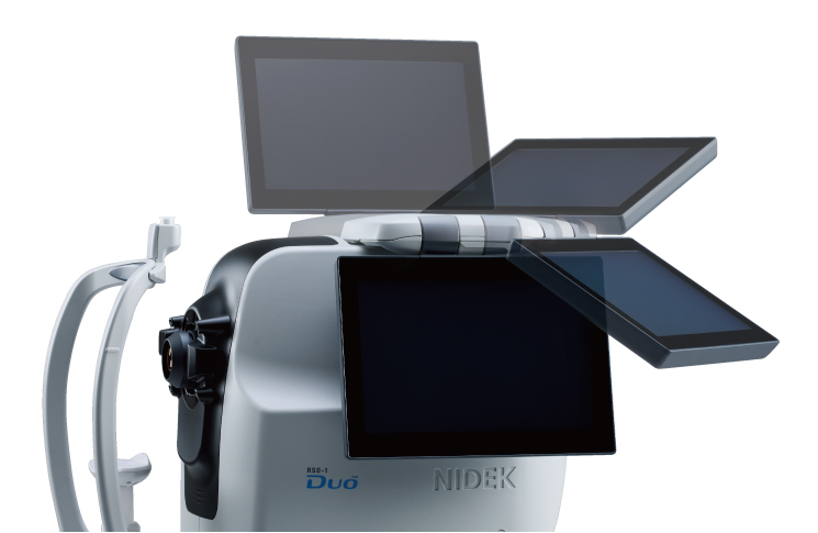
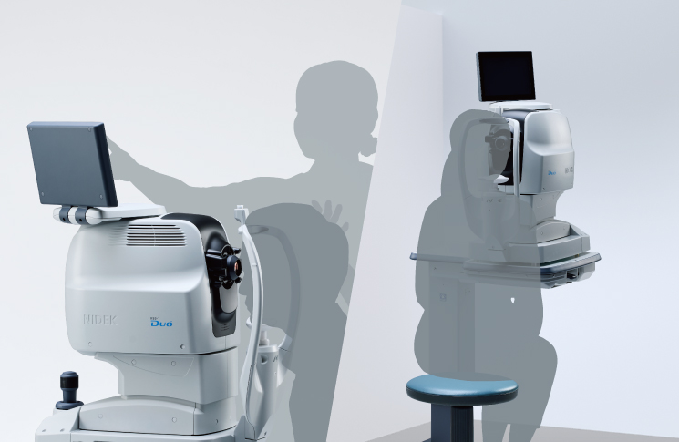
Space-saving designThe continuous range of tilt/swivel of the screen enables placement of the device anywhere in an examination room; installation against a wall or in a corner is now possible. |
 |
 |
Flexible data managementMore options for connectivity allow flexible data management. |
| 1. Standalone / Direct DICOM The RSD-1 Duo alone can provide basic analysis reports and print them out. Direct DICOM connectivity accommodates seamless data integration with PACS and EMR without an external PC. 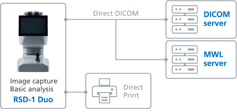 |
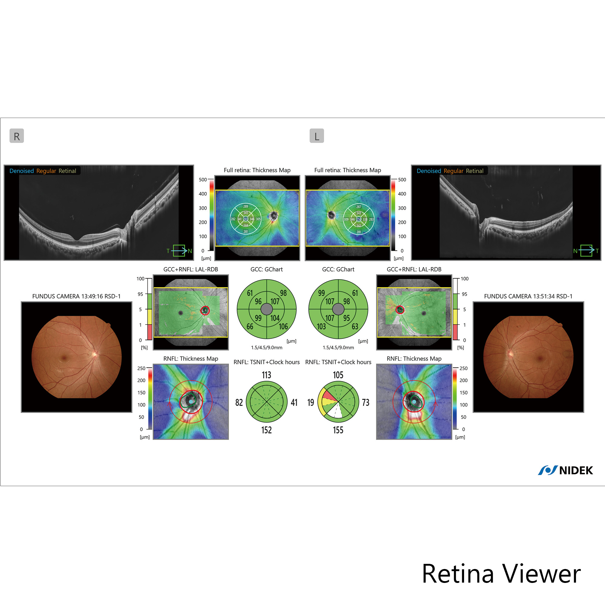
2. RSD-1 Duo + EyeLink / DICOM The images captured with the RSD-1 Duo can be automatically sent to EyeLink for advanced analysis. EyeLink stores all patient data on its server, enabling multiple PCs throughout the clinic to access the data, review images, and perform analysis using the dedicated viewer application, Retina Viewer. 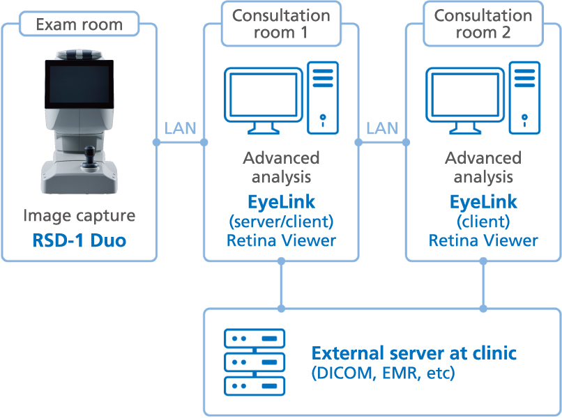 |
Downloads

Videos
Product Information
|
Related Products
Eye Care Data Management Platform
EyeLink™
Scanning Laser Ophthalmoscope
Mirante SLO/OCT
Mirante SLO
Optical Coherence Tomography
RS-1 Glauvas
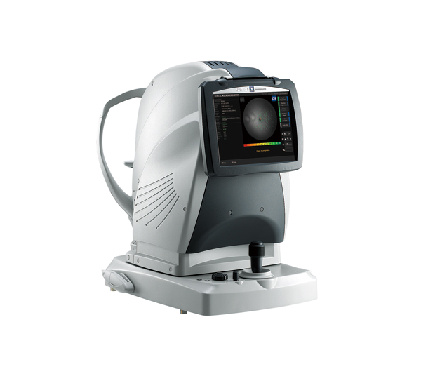
Microperimeter
MP-3
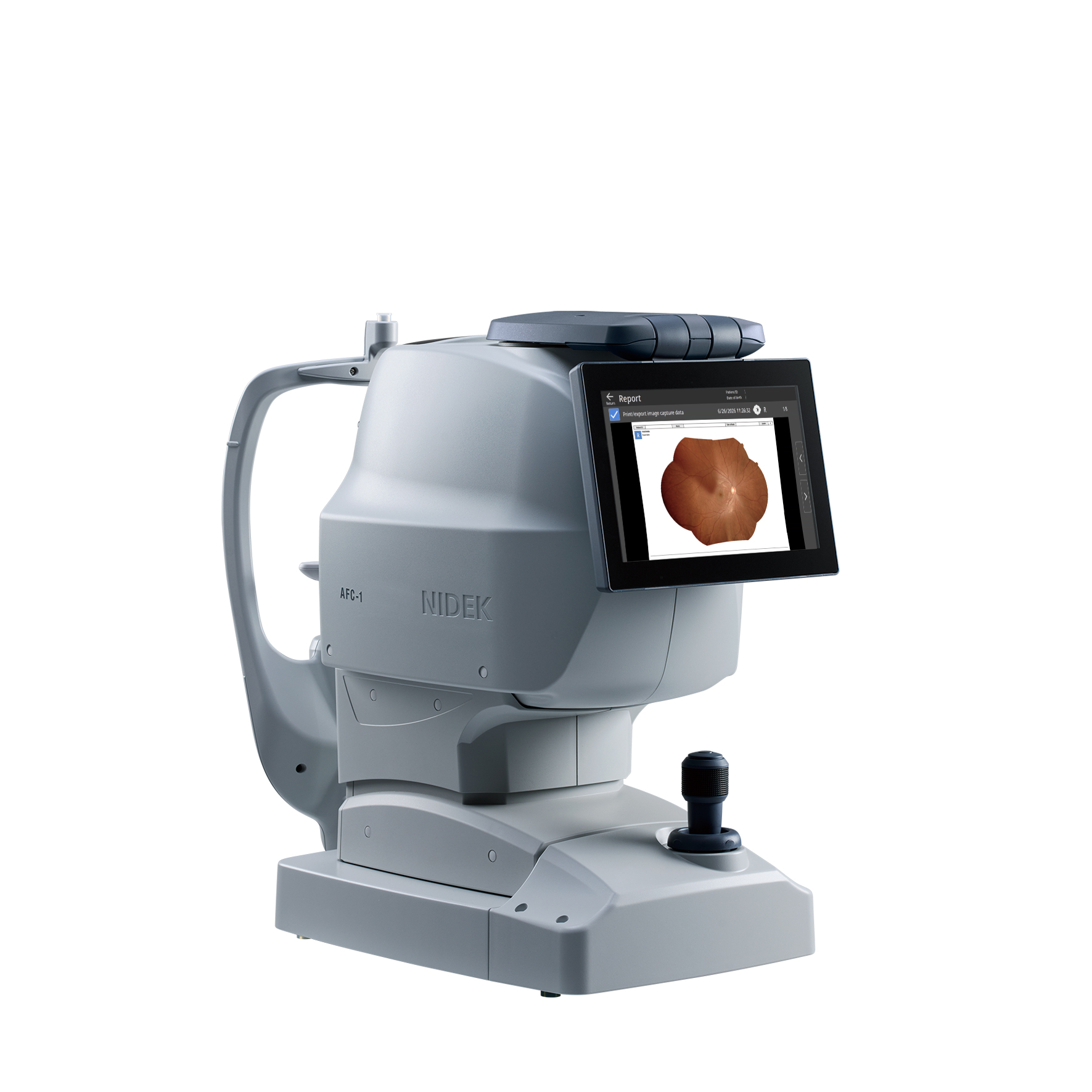
Non-mydriatic Auto Fundus Camera
AFC-1
NOTE
The availability of products differs from country to country depending on the status of approval.
Specifications and design are subject to change without notice.
- Product/model name
- Optical Coherence Tomography RSD-1