


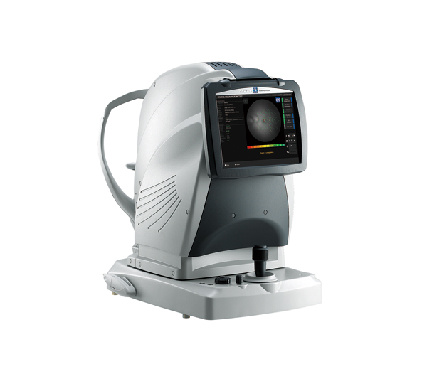
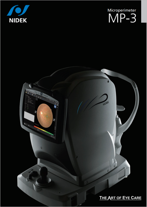
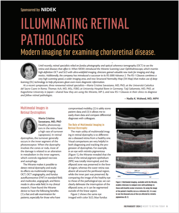
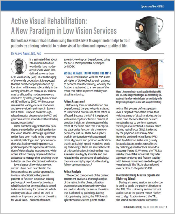
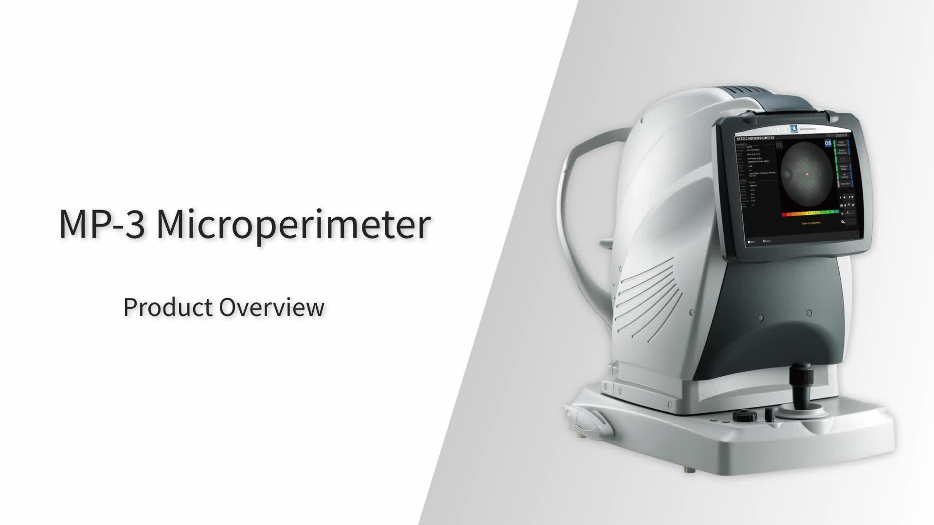
Microperimeter
MP-3
Features
- Microperimetry with a wide measurement range
- Fixation test with a precise tracking system
- High resolution non-mydriatic fundus camera
- Feedback exam for visual rehabilitation
- Scotopic microperimetry
- Auto tracking and auto alignment
Detailed Information
Microperimetry
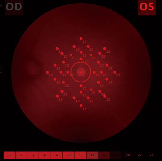
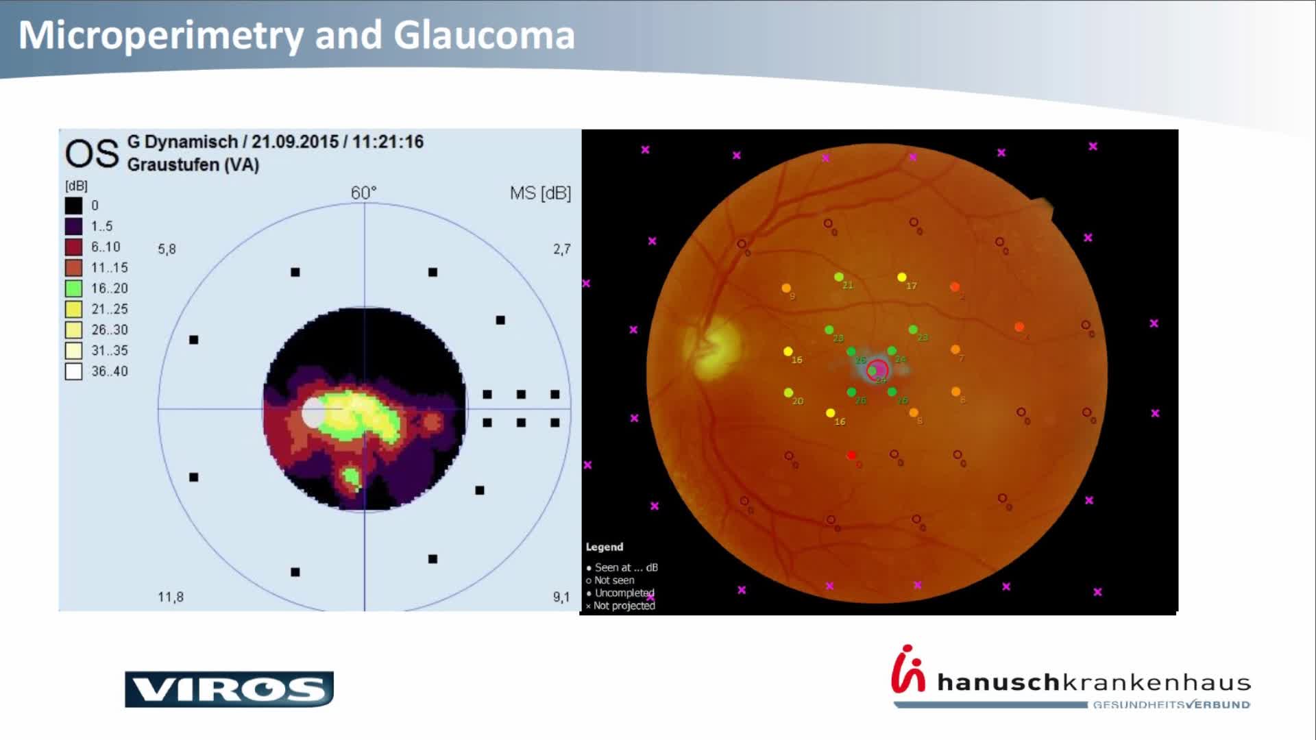
Wide measurement range
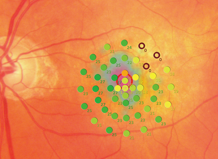
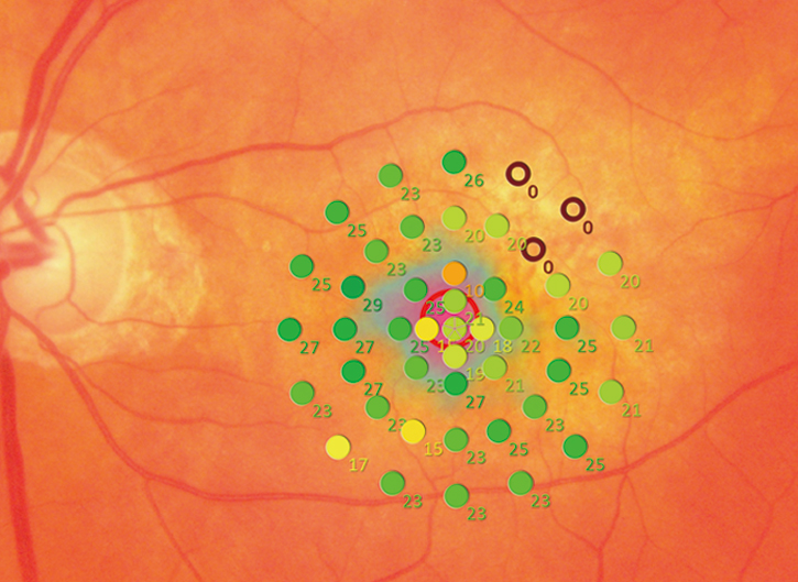
The MP-3 has a wider range of stimulus intensity, from 0 to 34 dB, compared to the MP-1. The MP-3 measures perimetric threshold values, even for normal eyes. A maximum stimulus luminance of 10,000 asb* allows evaluation of low-sensitivity.
*In accordance with ISO12866 measurement methods
MP-3 Normal Eye Image (34dB) |
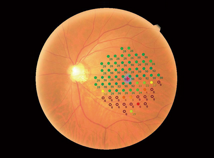
MP-3 Glaucomatous Eye Image (34dB) |
Fixation test
Precise tracking system
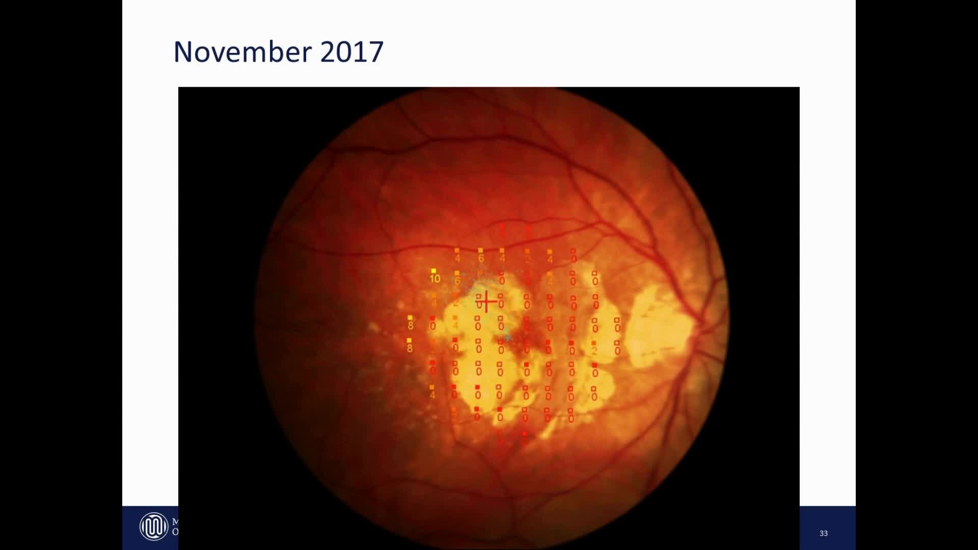
The MP-3 can measure fixation and determine the preferred retinal locus, simply by having the patient fixate on a target. Constant tracking of the eye during microperimetry allows evaluation of fixation in patients with central visual field defects and determines whether fixation improves after treatment.
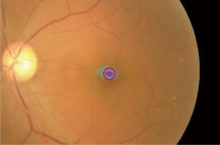
Stable Fixation |
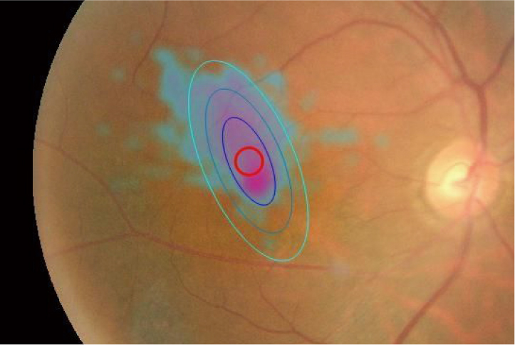
Unstable Fixation |
Retinography
High resolution non-mydriatic fundus camera
| An easy to use 12-megapixel fundus camera is incorporated into the MP-3 and acquires high resolution images of retinal pathology. | 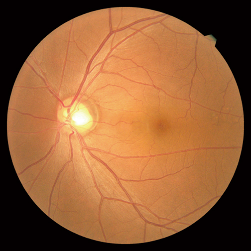
Fundus Camera Image |
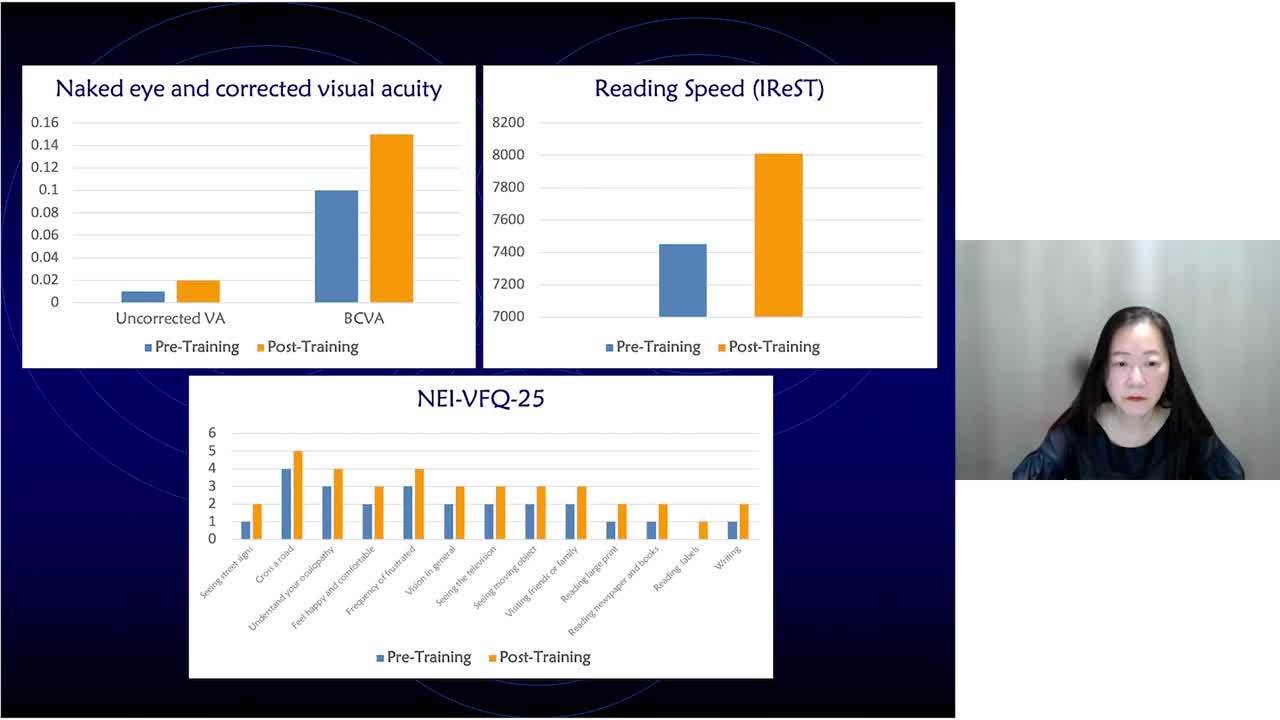
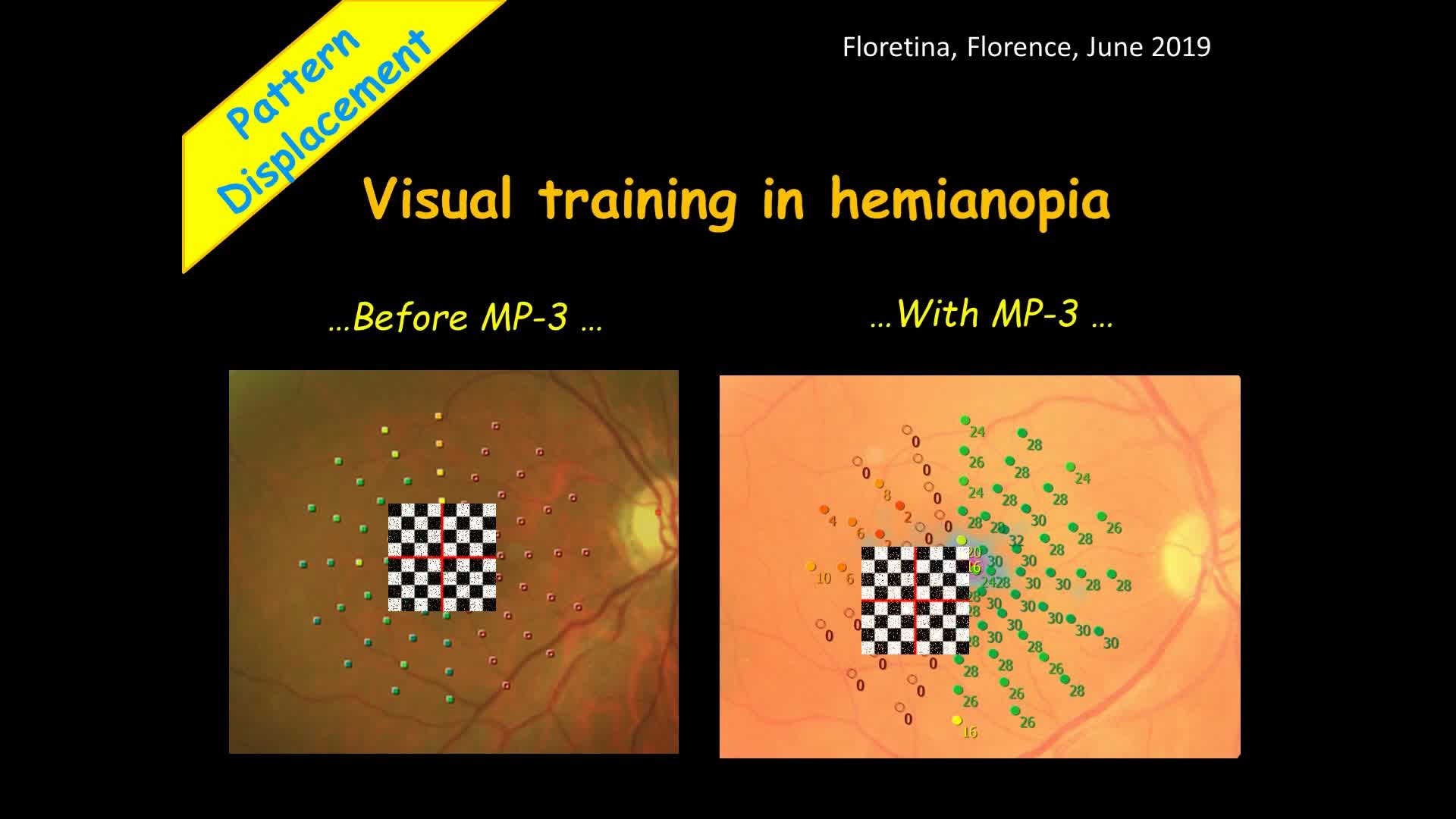
Feedback exam for visual rehabilitation
| The visual rehabilitation mode trains low-vision patients who have lost foveal fixation to relocate their preferred retinal locus (PRL) to a different region, called the trained retinal locus (TRL). The TRL is predetermined by a physician, and fixation rehabilitation allows the patient better functional vision (i.e. reading speed) due to increased fixation stability and visual outcomes.
Active flickering pattern stimulation and cheery music create an effective and pleasant training experience for the patient. |
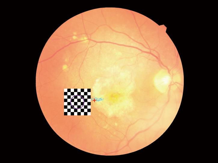 |
Image courtesy of the National Centre of Services and Research for the Prevention of Blindness and Rehabilitation of Visually Impaired – IAPB Italia Onlus, Rome – Italy
Related article
Active Visual Rehabilitation: A New Paradigm in Low Vision Services
By Filippo Amore, MD, PhD
https://www.nidek-intl.com/case_report/active-visual-rehabilitation-a-new-paradigm-in-low-vision-services/
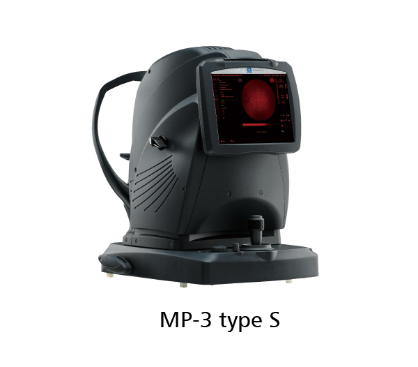
Scotopic microperimetry (available for the MP-3 type S)
| The MP-3 type S measures retinal functions under scotopic conditions (scotopic microperimetry) in addition to the standard functions of the MP-3.
Scotopic microperimetry is used to assess the changes in rod sensitivity of degenerative retinal diseases including age-related macular degeneration and some forms of retinitis pigmentosa. This modality can be used in clinical trials of new therapeutics for retinal diseases that impair rod function. |
MP-3 type S |
Auto tracking and auto alignment
| Auto tracking and auto alignment functions provide more accurate measurements increasing patient and operator comfort and efficiency. These functions allow easy follow-up and reduce variations between examiners, resulting in well-aligned follow up exams. |
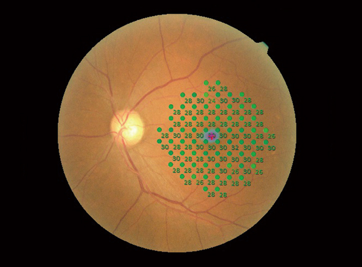
Region-specific test evaluation
| After completion of measurements, results can be evaluated in a specific region of interest to allow easier comparison with other pathology images. By specifying the region of interest, the average results in the region are displayed. | 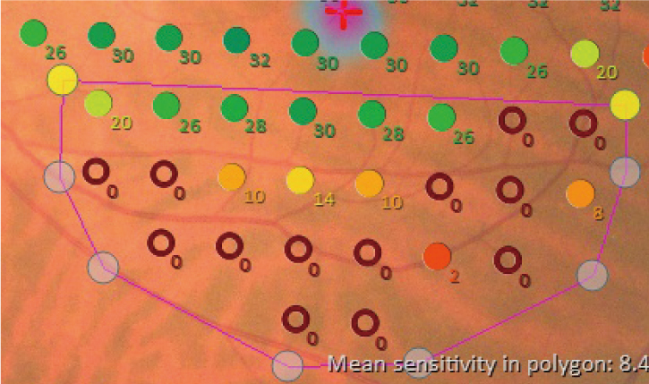
Magnified Image of Specified Fixation Point |
Follow-up test
| A follow-up test can be performed on the same area using the same parameters as a previous test. This feature allows evaluation of disease progression or assessment of pre- and post-treatment outcomes. Any differences in two microperimetry images are displayed for quick, intuitive interpretation. | 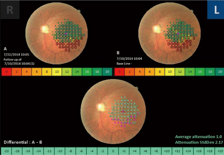
Follow-up Image |
Pre- and post-treatment comparison
Case of anti-VEGF treatment for age-related macular degeneration (AMD)
Pre-treatment |
Circle at 2° Percentage of fixation points 66.1% Circle at 4° Percentage of fixation points 92.1% Mean sensitivity: 20.4 |
Post-treatment |
Circle at 2° Percentage of fixation points 68.1% Circle at 4° Percentage of fixation points 95.5% Mean sensitivity: 20.9 |
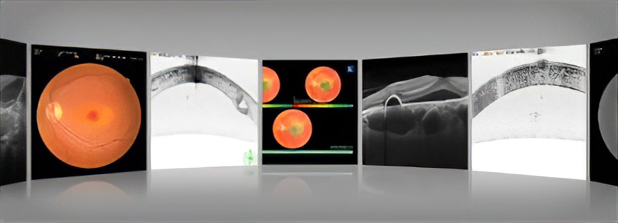
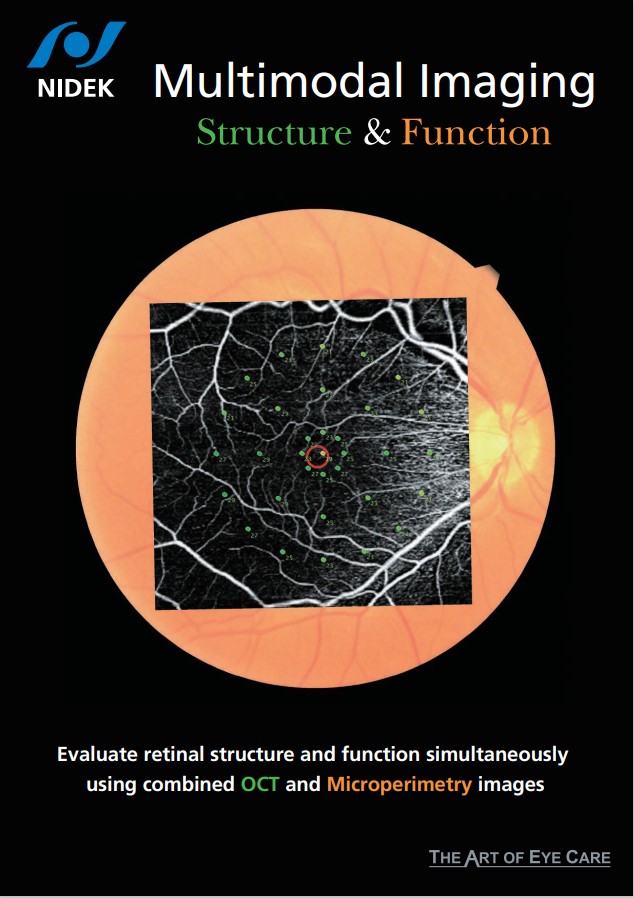
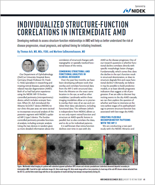
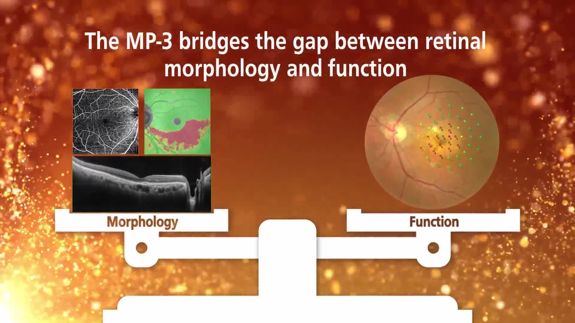
Structural and functional evaluation using OCT
Various OCT modalities captured by the Mirante can be registered with microperimetry.
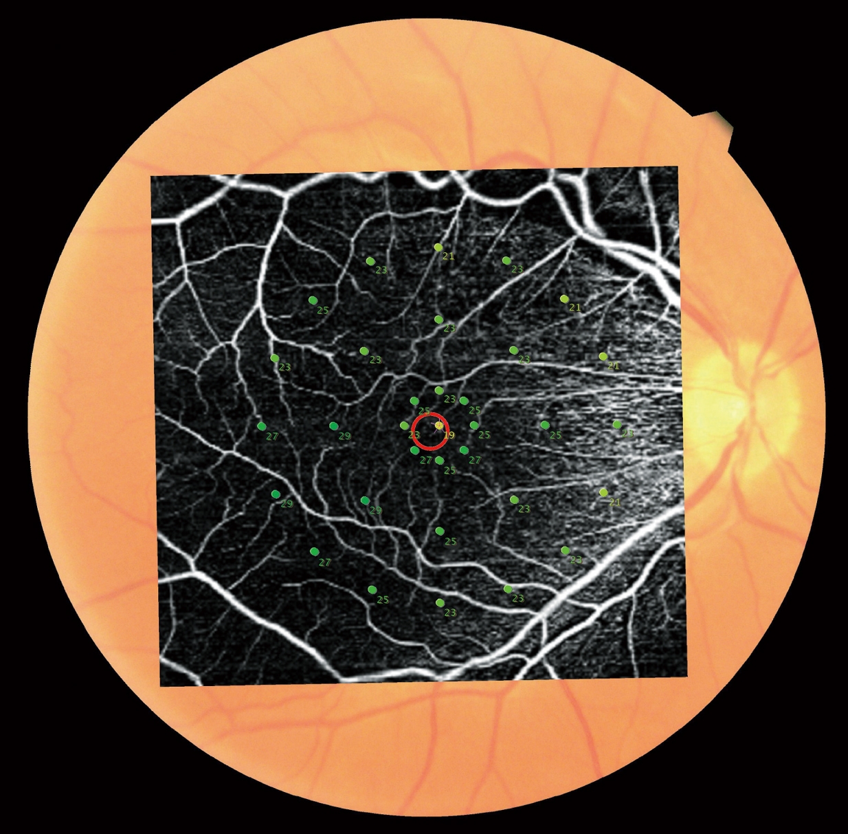
OCT Angiography+Microperimetry |

Normative DB+Microperimetry |
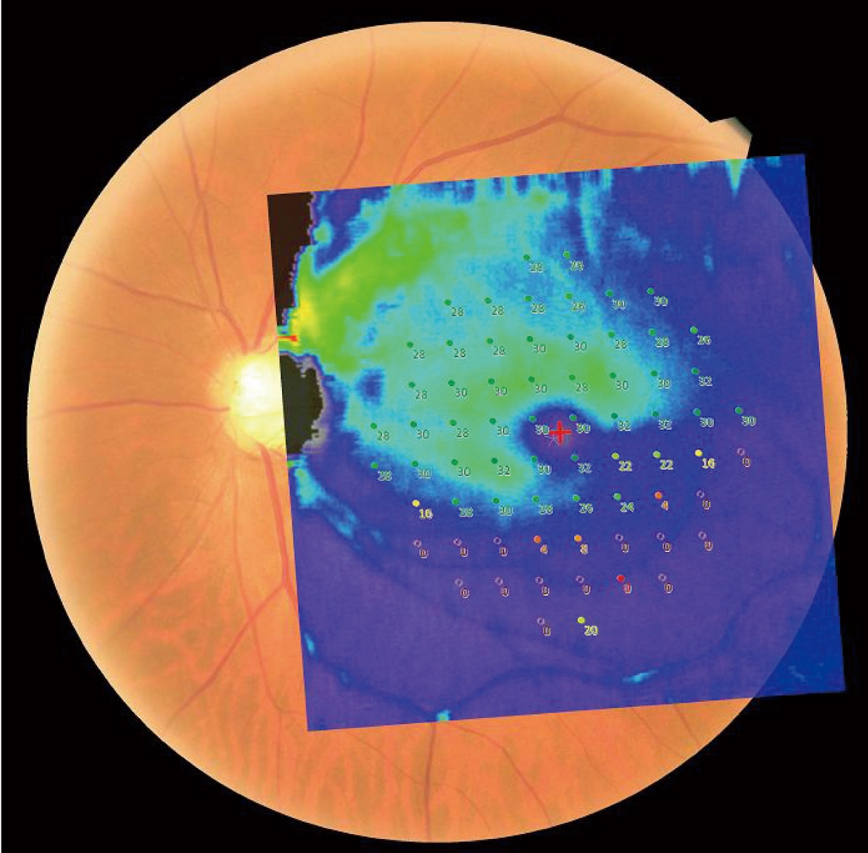
Thickness Map+Microperimetry |
Downloads


|
Videos


User Testimonials

Victor H. Gonzalez, MD
Gulf Coast Eye Institute, Valley Retina Institute, USA

Rodrigo Abreu Gonzalez, MD, PhD, FEBO
University Hospital of La Candelaria, Spain

Anna Tan, MBBS, M Med(Ophth), FRCS(Ed), FAMS
Singapore National Eye Centre, Singapore

Sadiq N Syed, MD
Maryland Retina, USA

Enzo Maria Vingolo, MD
University Sapienza of Rome “Polo Pontino” Department of Sense Organs, Italy

Thanapong Somkijrungroj, MD
Chief of Uveitis Unit, Retina Unit, King Chulalongkorn Memorial Hospital and University, Thailand

Laurentino Biccas Neto, MD, PhD
Ocular Vitória, Brazil

Thales A. C. de Guimarães, MD
Moorfields Eye Hospital NHS Foundation Trust, UK
UCL Institute of Ophthalmology, UK
Victor H. Gonzalez, MD
Gulf Coast Eye Institute, Valley Retina Institute, USA
Rodrigo Abreu Gonzalez, MD, PhD, FEBO
University Hospital of La Candelaria, Spain
Anna Tan, MBBS, M Med(Ophth), FRCS(Ed), FAMS
Singapore National Eye Centre, Singapore
Sadiq N Syed, MD
Maryland Retina, USA
Enzo Maria Vingolo, MD
University Sapienza of Rome “Polo Pontino” Department of Sense Organs, Italy
Thanapong Somkijrungroj, MD
Chief of Uveitis Unit, Retina Unit, King Chulalongkorn Memorial Hospital and University, Thailand
Laurentino Biccas Neto, MD, PhD
Ocular Vitória, Brazil
Thales A. C. de Guimarães, MD
Moorfields Eye Hospital NHS Foundation Trust, UK
UCL Institute of Ophthalmology, UK
Related Products
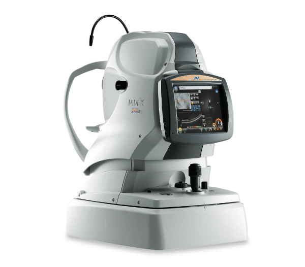
Scanning Laser Ophthalmoscope
Mirante SLO/OCT
Mirante SLO
Optical Coherence Tomography
RS-1 Glauvas
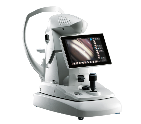
Optical Coherence Tomography / Fundus Camera
Retina Scan Duo™2
Gonioscope
GS-1
Ophthalmic YAG and SLT Laser System
YC-200 S plus
Ophthalmic YAG Laser System
YC-200
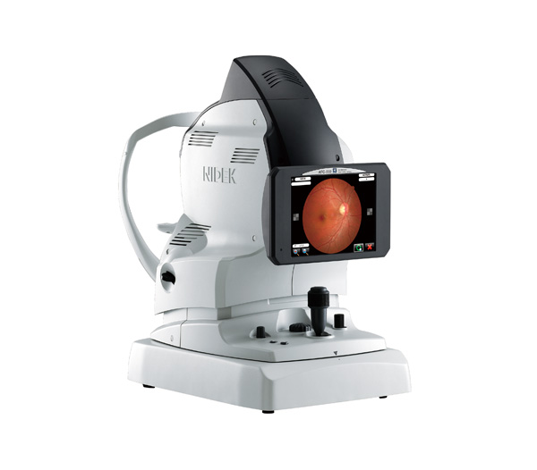
Non-mydriatic Auto Fundus Camera
AFC-330
NOTE
The availability of products differs from country to country depending on the status of approval.
Specifications and design are subject to change without notice.
- Product/model name
- Microperimeter MP-3