


































 /SC-1600

 TOP
TOP


Research and Development of Visual Prosthesis
“Invisible to Visible” — The development of visual prostheses has been THE goal of NIDEK since its founding.
Since 2001, NIDEK has been engaged in the research and development of an artificial retina system, which is a type of visual prosthesis that stimulates the retina.
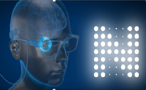
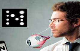
We aim to introduce an implantable medical device that restores visual perception as images composed of multiple points of light by electrically stimulating retinal nerve cells in individuals who have lost their sight.
Vision and the Artificial Retina System
■How Vision Works
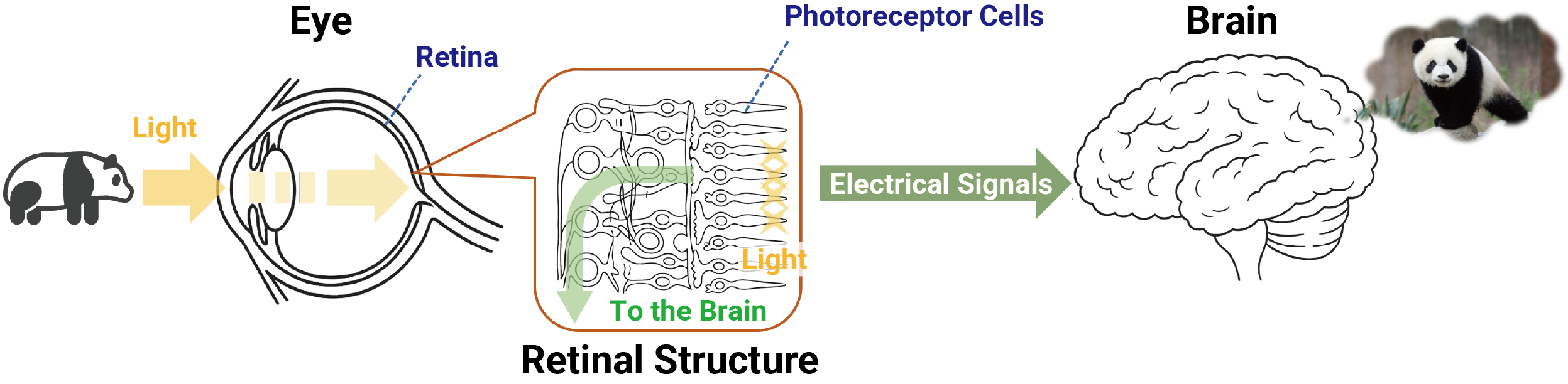
The retina plays an essential role in vision.
The retina, located at the back of the eye, is a very thin layer of neural tissue, approximately 0.2 mm thick. When light reaches the retina, it is converted into electrical signals which are transmitted to the brain through the optic nerve. The brain processes these signals, allowing us to perceive shapes, brightness, and colors.
There are various types of nerve cells within the retina, including photoreceptor cells that detect light. Photoreceptor cells contain light-sensitive substances that change their activity and generate electrical signals upon exposure to light. These signals are processed within the retina, transmitted through the optic nerve to the brain, and interpreted as visual information such as shapes, brightness, and colors.
■How the Artificial Retina System Works
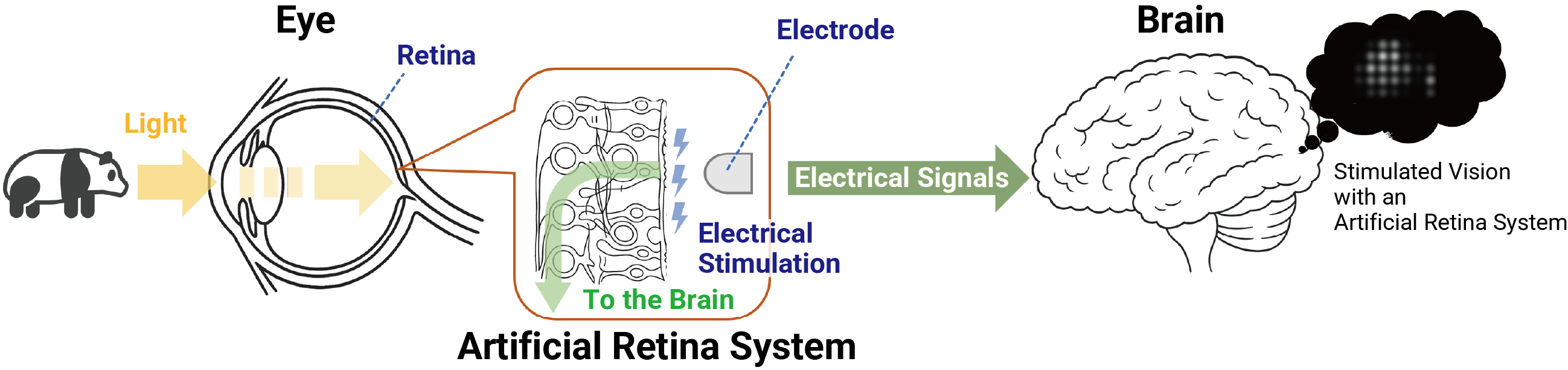
The Artificial Retina System consists of a camera and electrodes implanted inside the body.
Images captured by a camera mounted on a pair of glasses are converted into electrical signals and transmitted wirelessly to an implanted device. These signals are then delivered to electrodes implanted near the retina, stimulating the visual pathway and transmitting information to the brain.
Through this process, visual information can be perceived as cues of light and shape. However, the amount of information that can be conveyed is limited, and vision may be perceived as a collection of points of light.
The Artificial Retina System is a medical technology designed to supplement the retinal function that has been impaired by conditions such as retinitis pigmentosa.
■How Retinitis Pigmentosa Affects Vision
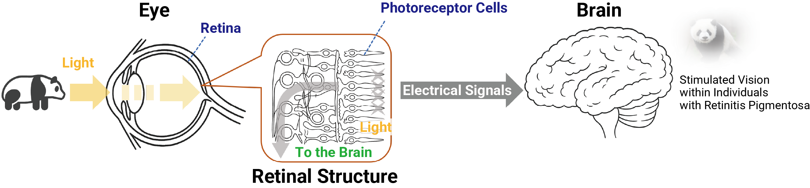
Retinitis pigmentosa (RP) is a disease in which the photoreceptor cells in the retina gradually lose their function, causing vision to slowly change over time.
As these light-sensing cells become less effective, the retina’s ability to convert light into electrical signals and transmit them to the brain becomes weaker.
Consequently, people with RP may have trouble seeing in low-light conditions, and their field of vision may gradually become narrower over time.
Although the rate of progression and changes in vision vary from person to person, the disease typically progresses slowly.
A characteristic feature of RP is that the gradual loss of photoreceptor cell function leads to gradual changes in vision.
NIDEK’s Artificial Retina System
■How NIDEK’s Artificial Retina System Works
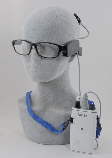
The Artificial Retina System consists of several components, including electrodes, a camera, a processing unit, and a wireless communication device.
The camera is mounted on a pair of glasses and images are captured based on the direction the user is facing. The processing unit, which contains a battery and signal-processing technology, is worn around the neck.
Images captured by the camera are converted into stimulation signals and transmitted through a wireless communication device to an implanted device in the head. The signals are then delivered from the implanted device to the electrodes placed inside the eye as electrical stimulation.
The electrical stimulation activates the retina, and the resulting signals are transmitted to the brain, where they are perceived as visual information such as brightness and shape.
■Implanted Device
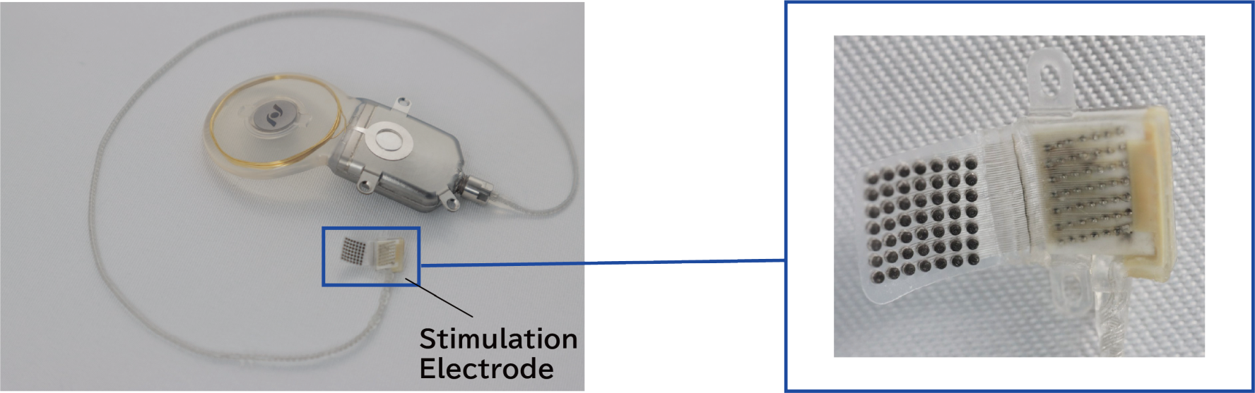
The implantable portion of the Artificial Retina System comprises a main unit with communication and control functions, along with an electrode array that stimulates the retina. A thin lead wire connects the main unit and the electrode array.
The main unit is composed of a circular coil approximately 30 mm in diameter connected to a 20 mm × 25 mm titanium case.
The electrode array measures 7 mm × 7 mm and contains 49 electrodes arranged in a grid pattern.
■Electrode Implantation
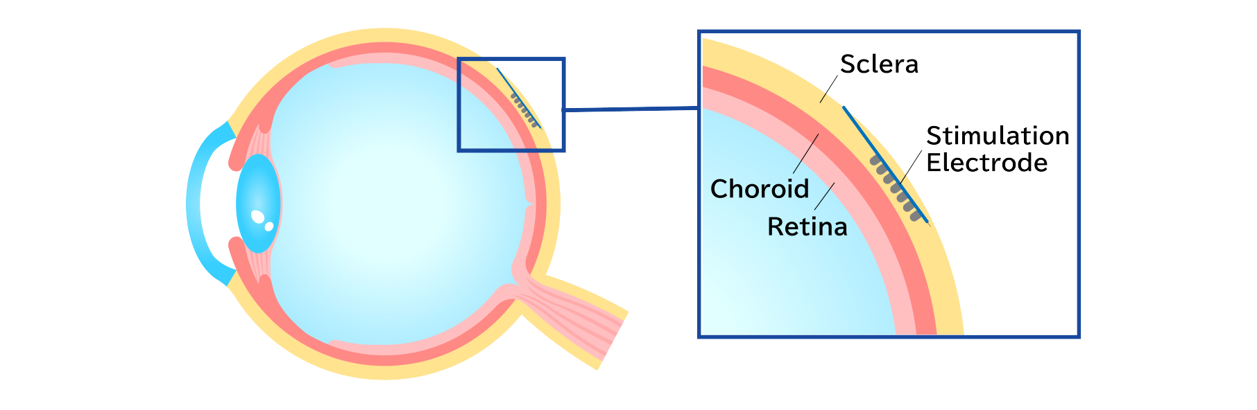
The electrode array of the Artificial Retina System is secured to the tissue surrounding the eyeball, known as the sclera.
This approach, known as the Japanese method, is considered to offer a high level of safety.
History of NIDEK’s Artificial Retina System Development
2001
NIDEK established the first Vision Institute for visual prostheses in Japan.
2001–2005
NIDEK has been entrusted by the New Energy and Industrial Technology Development Organization (NEDO Japan) to be a member in collaboration with several universities to promote research and development on visual prostheses as a national project of Japan. *1
*1: Basic research has been conducted in cooperation with the following research institutions and the NIDEK.
1) Nara Institute of Science and Technology, 2) Kyusyu University
2006–2007
This continuing project has been supported by the NEDO grant for the Application of Industrial Technology Innovation. It was carried out as a joint effort of the Ministry of Economy, Trade, and Industry, and the Ministry of Health, Labor, and Welfare. Clinical research is being conducted at Osaka University.
2010
Semi-chronic clinical research*2 was performed on patients with retinitis pigmentosa at Osaka University.
*2: Clinical research on implanting the Artificial Retina System in the human body for a longer period than that for acute clinical research (approximately a month).
2014–2015
Chronic clinical research*3 was performed on patients with retinitis pigmentosa at Osaka University.
*3: Clinical research on implanting the system in the human body for approximately one year.
2016
Started device development for clinical trial.
2026
Initiated Japan’s first company-sponsored clinical trial.