


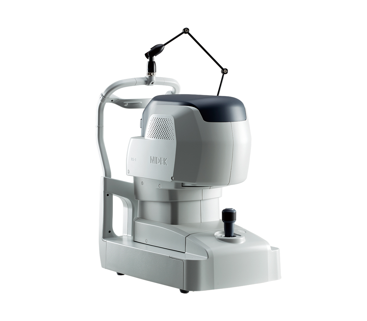
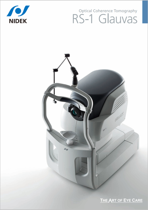
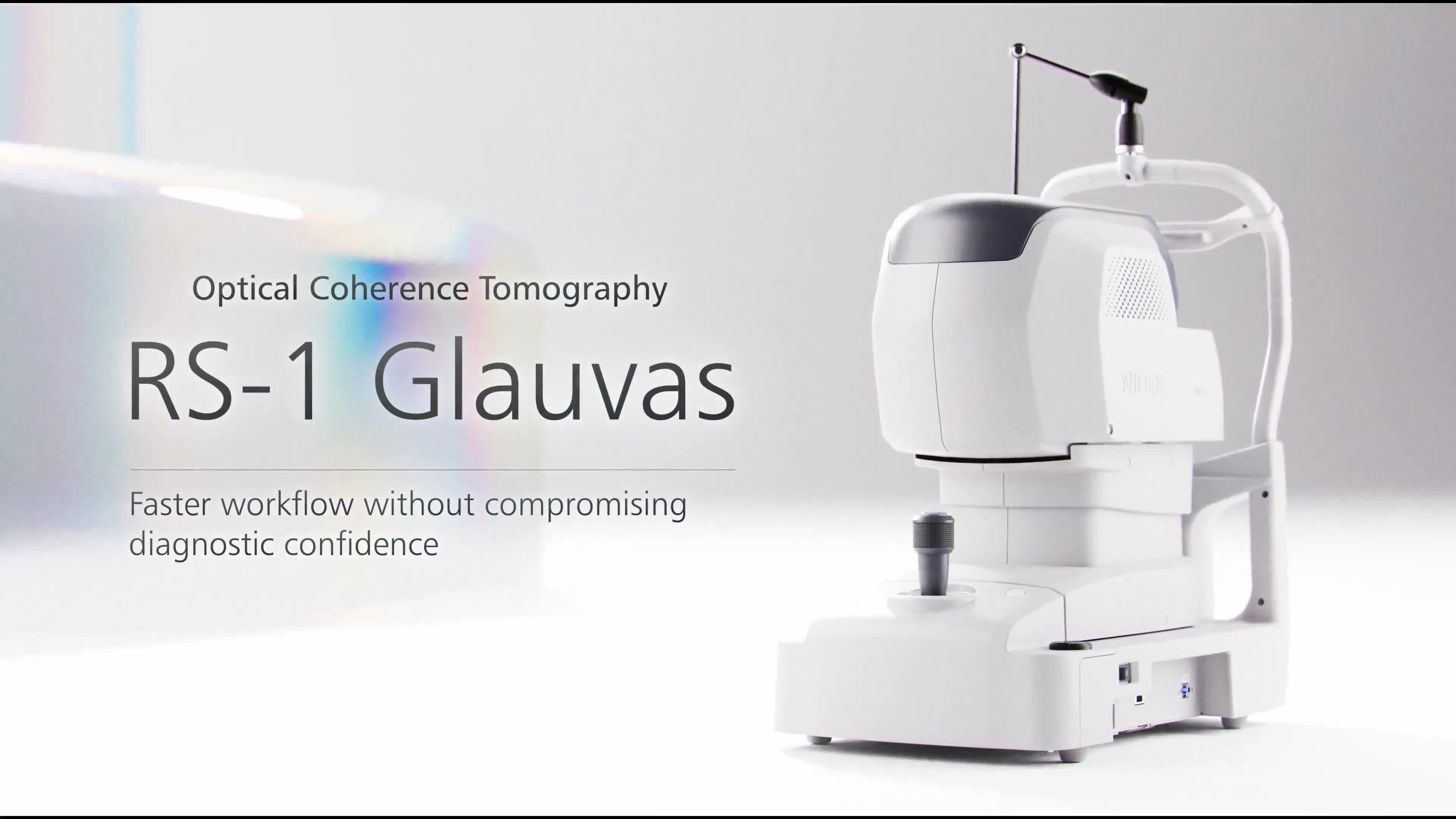
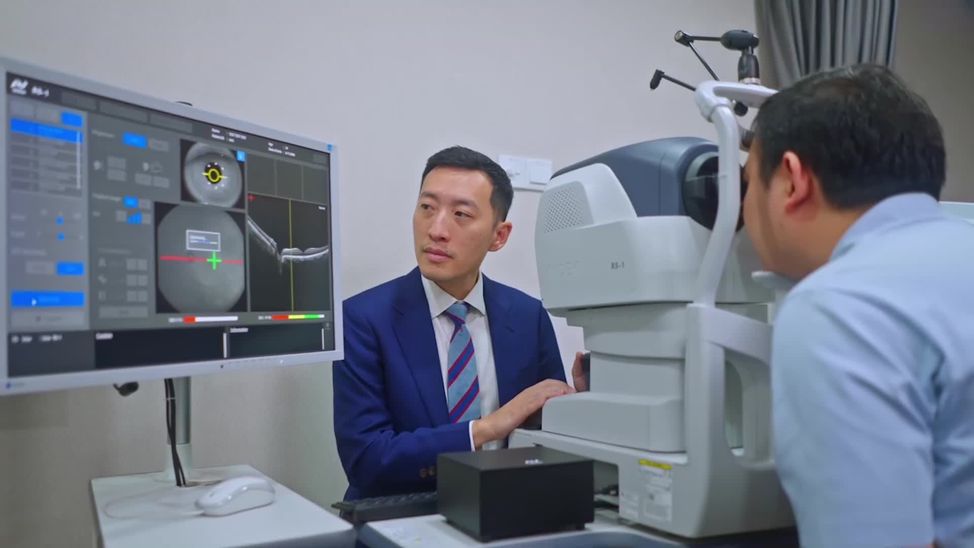
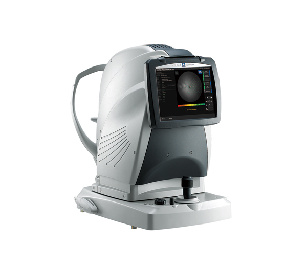
Optical Coherence Tomography
RS-1 Glauvas
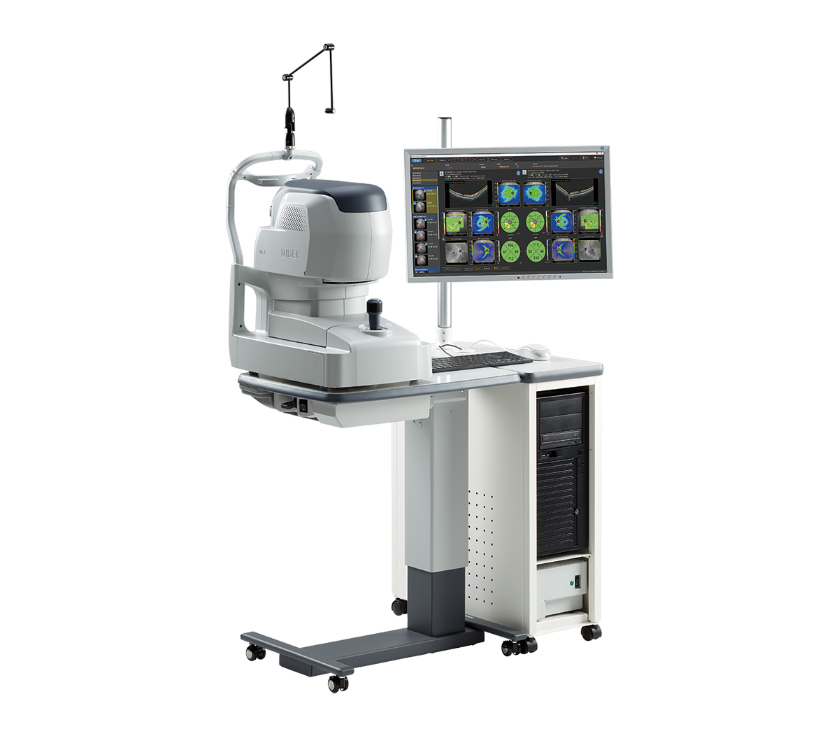
Features
- 250,000 A-scans/s high-speed imaging
- Wide, deep, high-resolution imaging
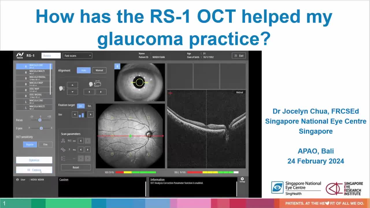
- Effortless operation and interpretation
- Advanced analytics
Detailed Information
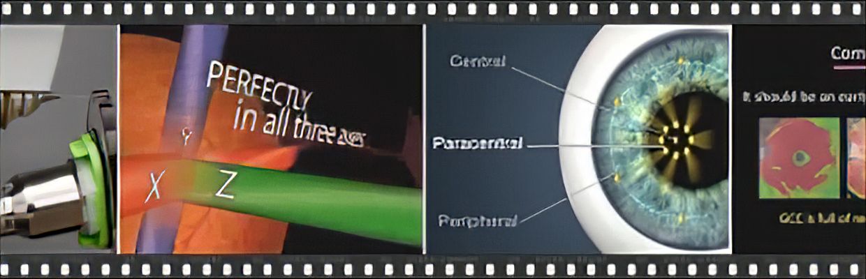
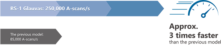
250,000 A-scans/s high-speed imaging
| The incorporation of 250,000 A-scans/s accelerates your workflow by reducing capture time. The high-speed imaging also addresses patient fixation errors thus contributing to greater image clarity and patient comfort.
|
 |
 |
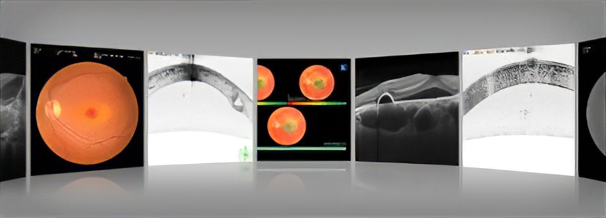
Wide, deep, high-resolution imaging
With RS-1 Glauvas, a single B-scan image clearly presents the area from the optic nerve head to the temporal vascular arcade, and the 4.2 mm depth B-scan imaging readily captures the oblate retinal shape of myopic eyes. Improvements in AngioScan OCT-Angiography include wider and clearer images for assessing chorioretinal microvasculature.
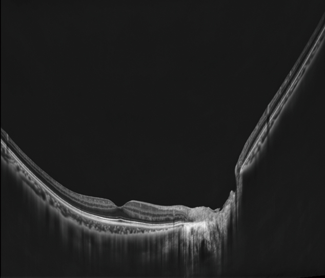
Macula line 16.5 mm / Scan depth 4.2 mm |
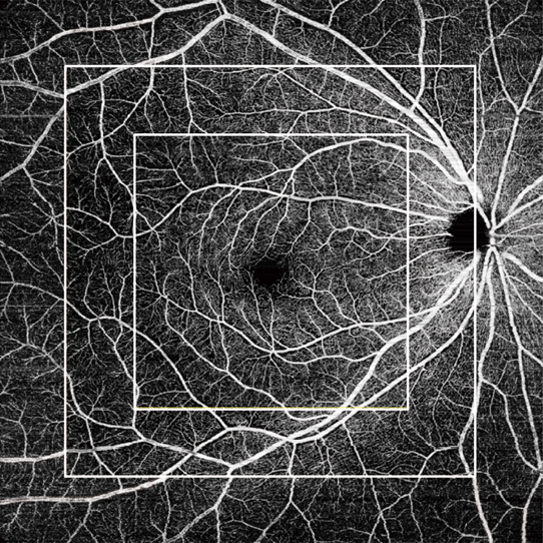
OCT-Angiography 6 x 6, 9 x 9, 12 x 12 mm |
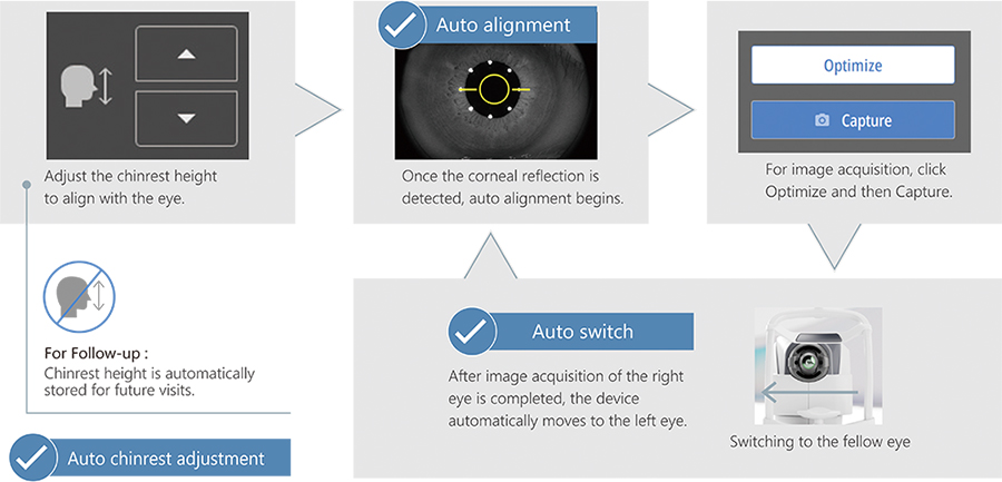
Effortless operation and interpretation
Advanced analytics
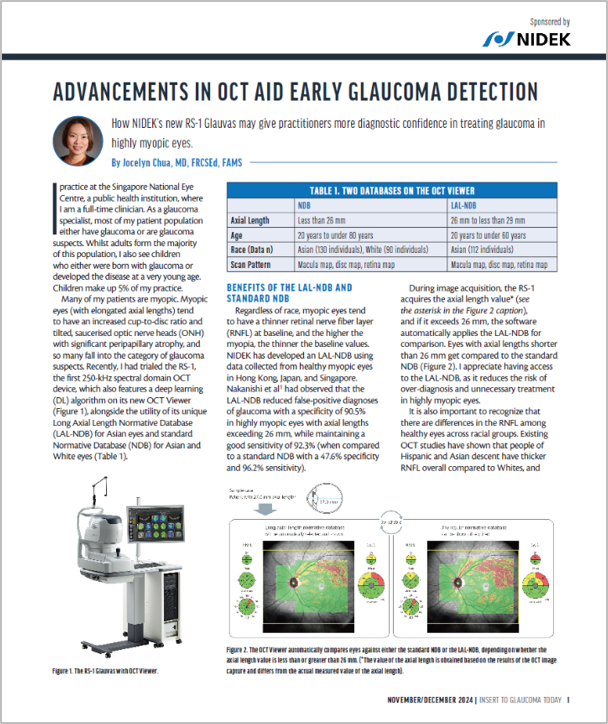
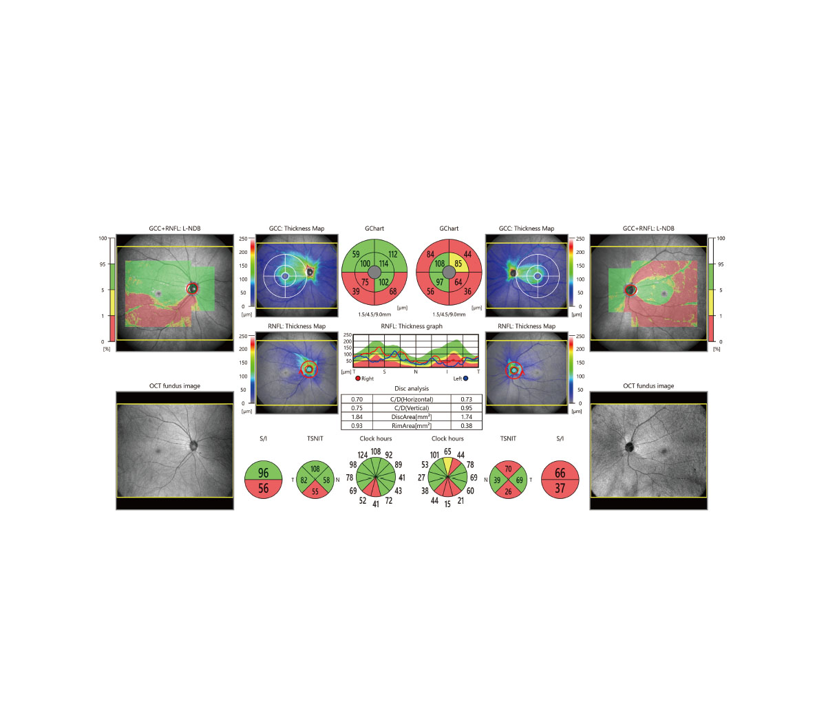
Glaucoma analysis in myopia
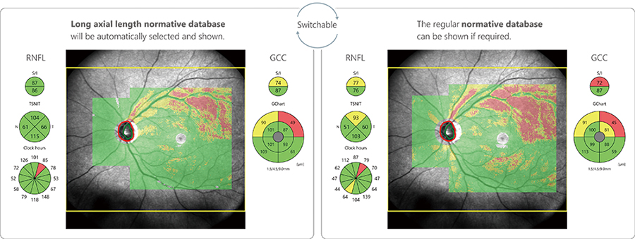
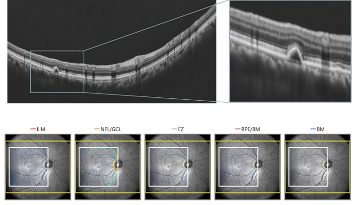
Less false positives with deep learning segmentation (DL segmentation)
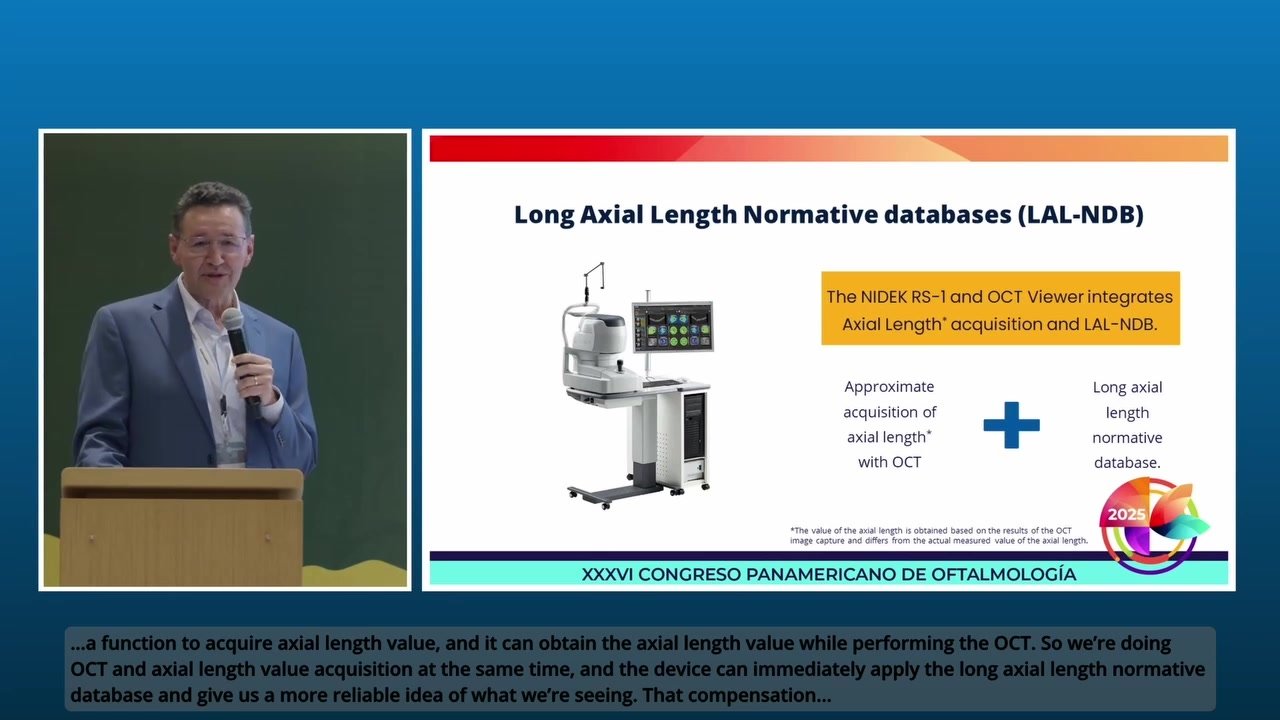
| The accuracy of segmentation affects the outcomes of glaucoma analysis. DL segmentation reduces artifacts and errors in the normative database and thickness maps even in eyes with opacities, thus decreasing false positives and enhancing clinic efficiency by reducing unnecessary follow-up visits. Additionally, the scan width correction allows precise analytics based on the patient’s axial length*2. | 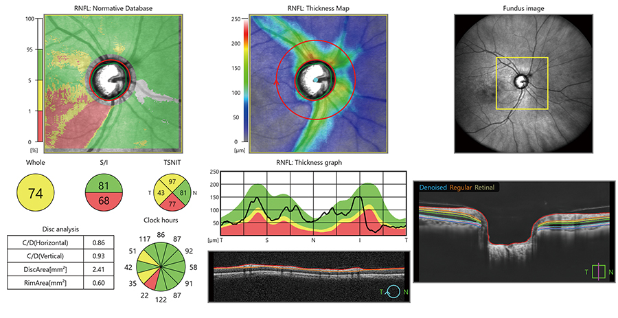 |
*1 Data was collected from a sample of Asian patients.
*2 The value of the axial length is obtained based on the results of the OCT image capture and differs from the actual measured value of the axial length.
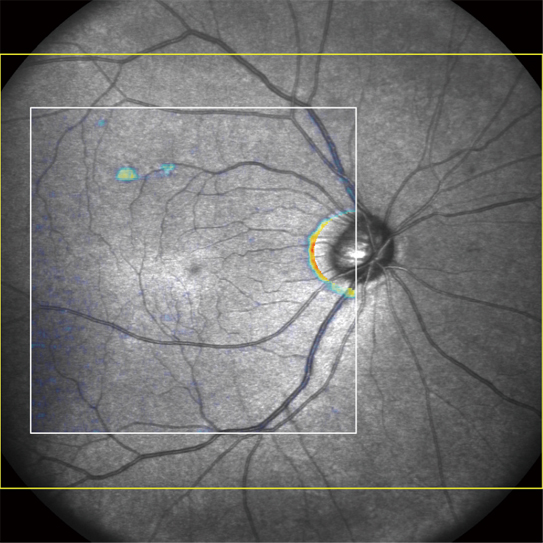
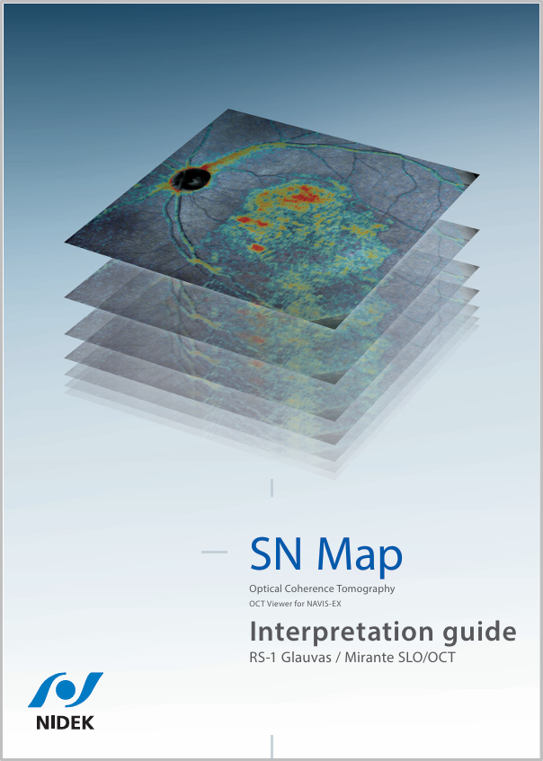
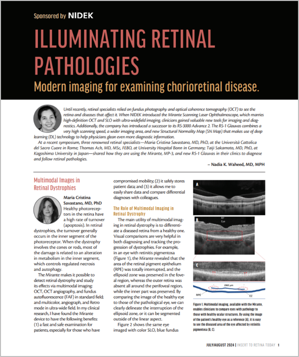
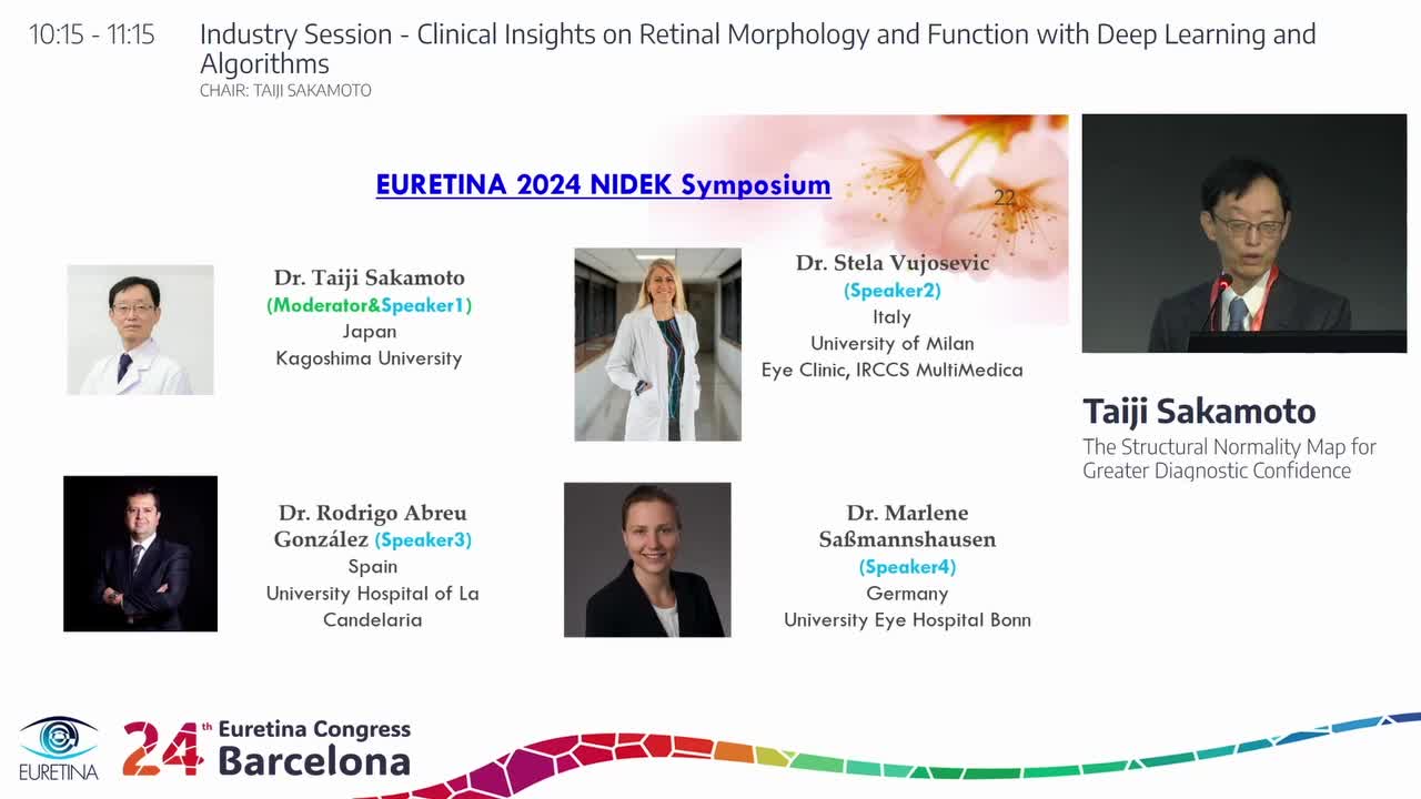
Structural Normality Map (SN Map) improving diagnostic confidence on early detection
| The DL segmentation provides an SN Map that presents structural abnormalities and changes. This functionality aids clinicians in detecting minute structural changes at a glance, contributing to greater diagnostic confidence even for early signs of retinal changes. | 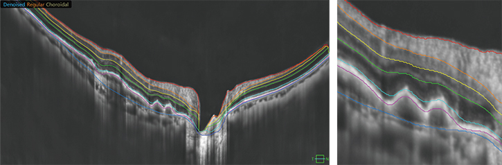
Less affected by a lesion or image contrast, the DL segmentation detects the edge of each layer on a B-scan image. Based on this highly precise segmentation, the SN Map can show subtle changes.
|
 |
 |
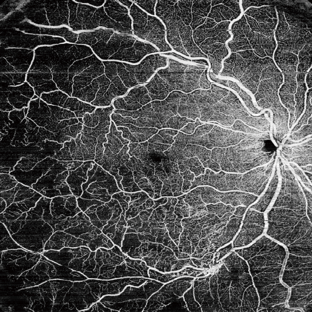
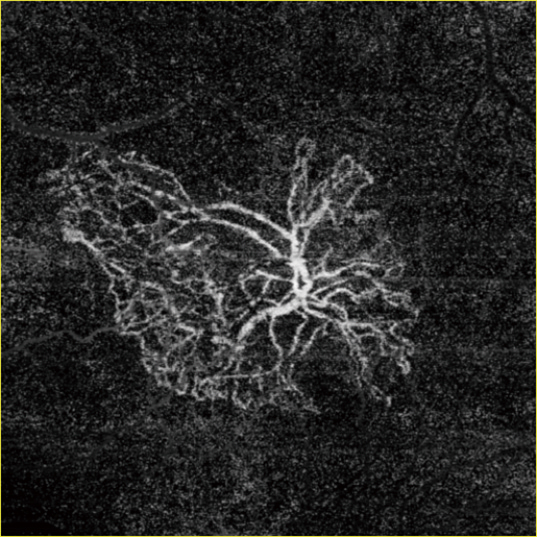
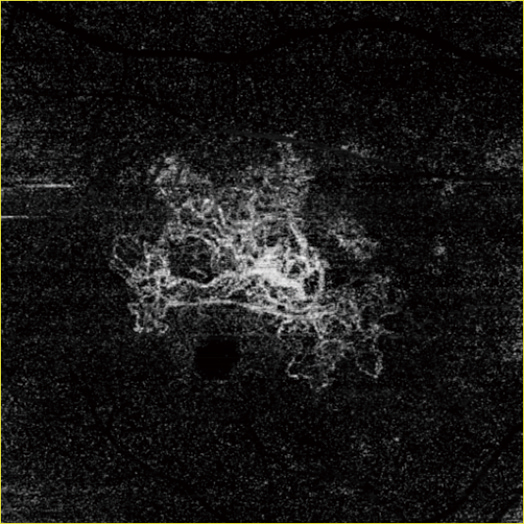
Enhanced OCT-A resolution with a new imaging algorithm
Sharp, wide OCT-Angiography enables detailed imaging of microvasculature even at the periphery of the scan area. In addition, a new algorithm, Complex OCT signal Difference Analysis Angiography 2 (CODAA 2), improves contrast, allowing for enhanced imaging. The high-definition OCT-Angiography enables greater clinical use of noninvasive imaging of retinal vascular diseases, reducing dependence on contrast dye tests.
 |
 |
 |
Downloads

Videos
Product Information
|

Introducción del producto (Spanish)
|
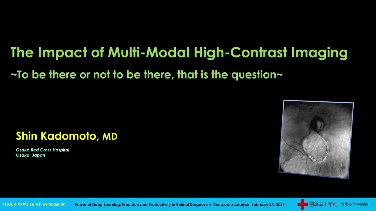
Lectures
|
|

|
|
|||
|
|
|
|
 EURETINA 2023 EURETINA 2023
|
User Testimonials
Kelvin Teo, MBBS, PhD —User Testimonial of RS-1
|
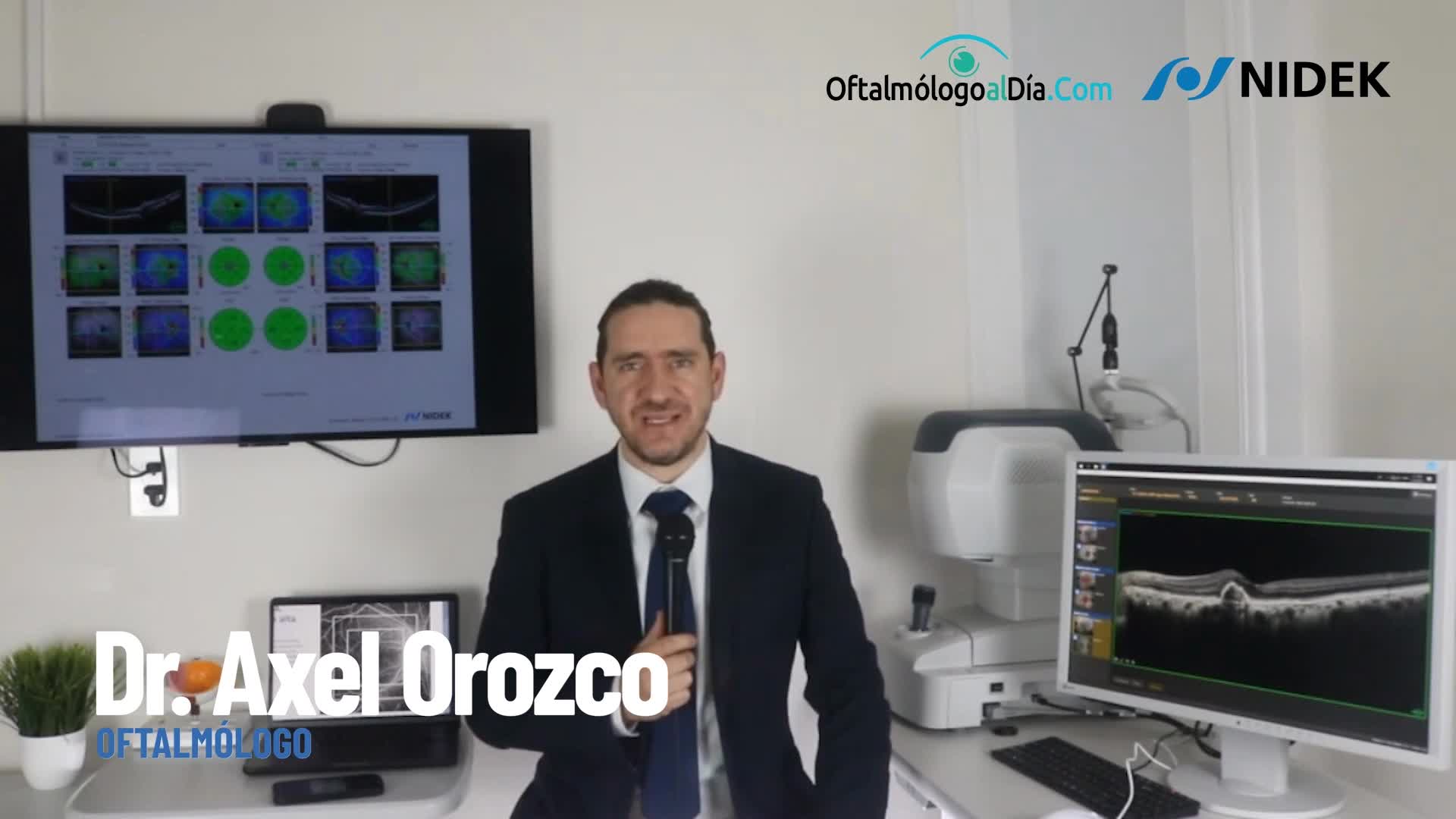
El Dr. Axel Orozco Hernández comparte su experiencia con el OCT RS-1 Glauvas (Spanish)
|
Related Products
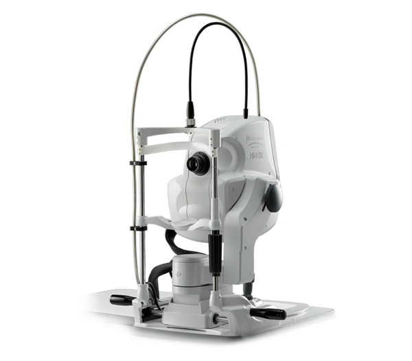
Scanning Laser Ophthalmoscope
Mirante SLO/OCT
Mirante SLO
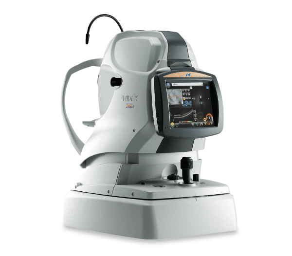
Optical Coherence Tomography / Fundus Camera
Retina Scan Duo™2
Software for NIDEK OCT series
Long Axial Length Normative Database
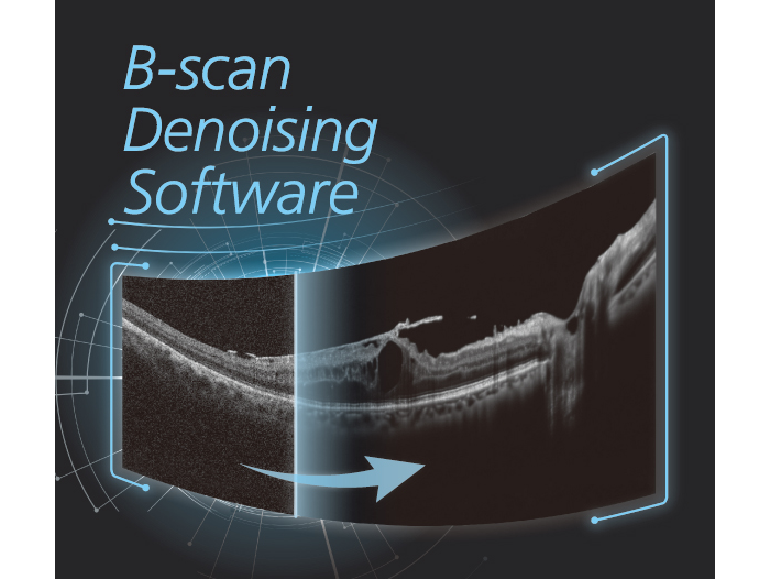
B-scan Denoising Software
for NIDEK OCT series
Microperimeter
MP-3
Gonioscope
GS-1
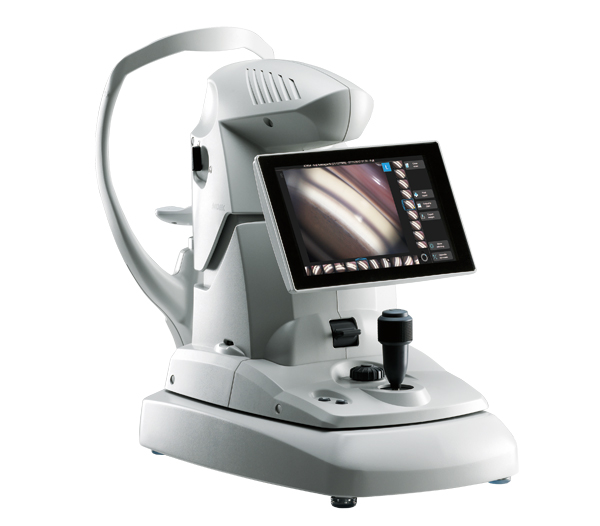
Non-mydriatic Auto Fundus Camera
AFC-330
NOTE
The availability of products differs from country to country depending on the status of approval.
Specifications and design are subject to change without notice.
- Product/model name
- Optical Coherence Tomography RS-1