

























 /SC-1600

 TOP
TOP


NIDEK launches the RSD-1 Duo™ OCT / Fundus Camera, AFC-1 Fundus Camera, and EyeLink™ Eye Care Data Management Platform
2026.09.03
| -3 September 2026, Gamagori, JAPAN- |
| NIDEK CO., LTD., a global leader in the design, manufacture, and distribution of ophthalmic, optometric, and lens edging equipment, is pleased to announce the launch of the RSD-1 Duo OCT / Fundus Camera, AFC-1 Non-mydriatic Auto Fundus Camera, and EyeLink Eye Care Data Management Platform. |
| RSD-1 Duo and AFC-1 | ||
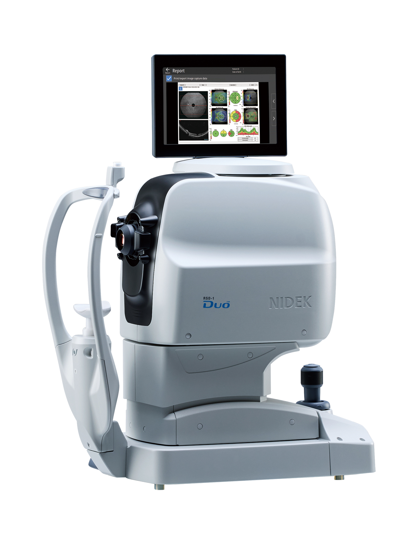
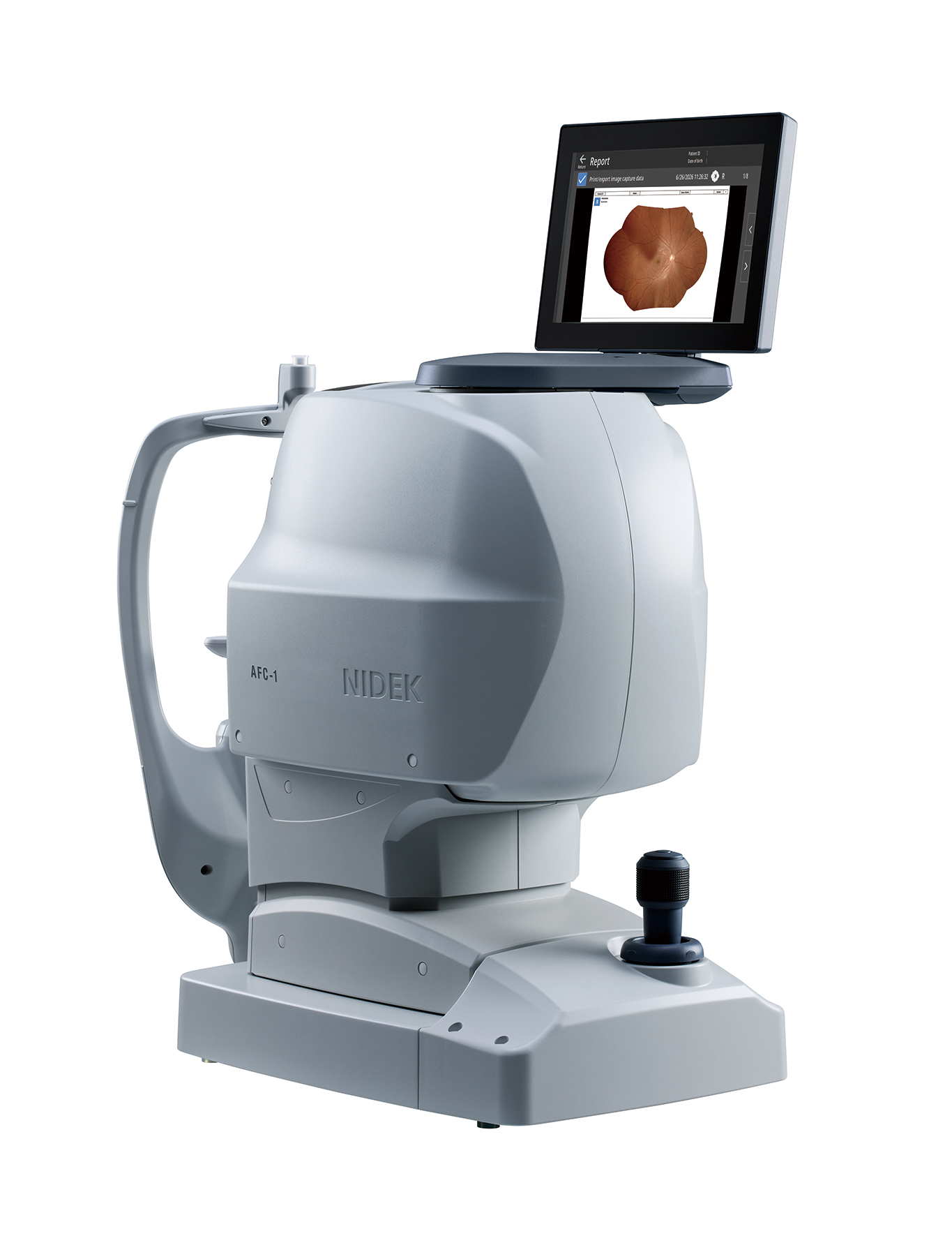
| The RSD-1 Duo, a combined OCT and fundus camera system, and the AFC-1 fundus camera, both increase efficiency across the entire clinical workflow, from image capture and screening to data management. Each device is highly automated, with intuitive operation that enables fast, consistent image capture, reducing operator workload and increasing patient comfort. |
|
|
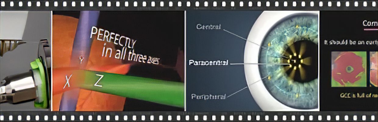
| For color fundus imaging, both devices feature advanced slit-scan technology delivering high-contrast, high-definition images with fewer artifacts and shadows. This technology also achieves higher imaging success rates and consistent 50° wide imaging even in small pupils (min. ø2.0 mm). Seamless auto-composite panorama imaging delivers stunning clarity, far surpassing the previous models. Panorama composition can be completed quickly on the device by automatically combining up to seven shots. Fundus autofluorescence is available on the FAF model of the RSD-1 Duo. | ||
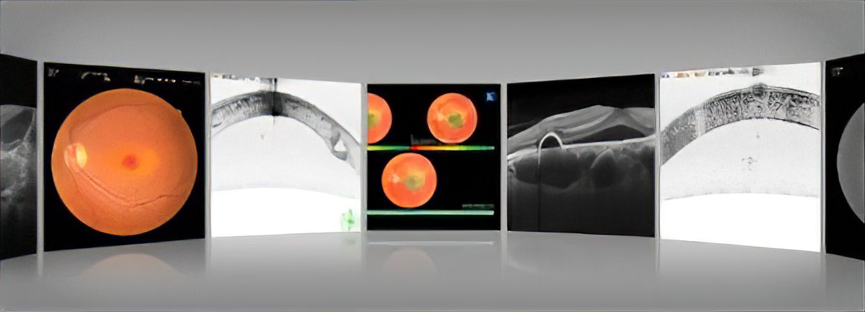
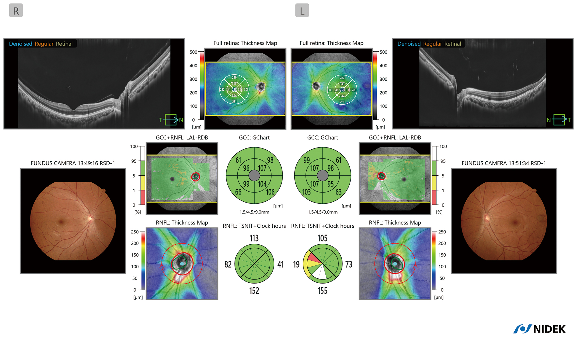
| For OCT imaging on the RSD-1 Duo, the B-scan image size has been expanded from the previous model, up to 16.5 mm in width and 3.0 mm in depth, capturing the area from the optic nerve head to the temporal vascular arcade and the oblate retinal shape of myopic eyes. The denoising technique for B-scan imaging provides high-definition images. Optional OCT-Angiography utilizes an advanced imaging algorithm to suppress artifacts and enhance vascular detail, improving visualization of clinically relevant information. Without the need for an external PC, the device can provide basic analysis and reports. The retina map, capturing both the disc and macula, enables reference database (RDB)*1 analysis for both the RNFL and GCC on a single OCT image, facilitating efficient diagnostic screening. For glaucoma analysis in axial myopia, the long axial length reference database (LAL-RDB)*2 presents analysis with axial length compensation. The RSD-1 Duo automatically selects the optimal database from either the regular RDB or LAL-RDB by acquiring and using the axial length*3 which is a parameter for scan width correction. | ||
| With flexible installation, PC-free operation, and expanded connectivity, including direct DICOM connectivity and EyeLink, the RSD-1 Duo and AFC-1 adapt seamlessly to a wide range of clinical environments and workflows. | ||
| EyeLink | ||
| The images captured with the RSD-1 Duo and AFC-1 can be automatically sent to EyeLink, when connected, which stores all patient data on its server, enabling multiple PCs throughout the clinic to access the data and review images. | ||
| Additionally, Retina Viewer, an optional dedicated viewer application, performs advanced analysis on the OCT and OCT-A images. Less affected by a lesion or image contrast, the deep learning segmentation (DL segmentation) detects the edge of each layer on a B-scan image. Based on this highly precise segmentation, the unique Structural Normality Map (SN Map) detects and highlights structural abnormalities, including subtle retinal changes, facilitating rapid interpretation. All B-scan and OCT-A images can be denoised using optional denoise licenses. OCT-A quantitative analysis functions provide metrics to assess disease progression and treatment effects. Follow-up analysis with automatic image registration allows clinicians to review chronological changes at the same location even on protocols with different scan sizes and/or orientations. ACA analysis, pachy map, and epithelium map allow thorough analysis of the anterior segment. |
|
|
| “We believe that the RSD-1 Duo and AFC-1, featuring effortless image capture, high-quality imaging, PC-free operation and analysis, and broad connectivity, streamline clinical workflows and support faster, more confident decision-making.” says Motoki Ozawa, President and CEO of NIDEK CO., LTD. “EyeLink delivers a dedicated viewer with the advanced analysis capabilities that clinicians demand, and we will continue to expand the platform to surpass their expectations.” |
| NIDEK will unveil these three products in person at ESCRS 2026 in London, UK (11–14 September). Visitors are invited to experience these new technologies firsthand at NIDEK booth D.300. |
| *1 Data was collected from a sample of Caucasian and Asian patients. *2 Data was collected from a sample of Asian patients. *3 The value of the axial length is obtained based on the results of the OCT image capture and differs from the actual measured value of the axial length. |
| Product/model name: Optical Coherence Tomography RSD-1 Class IIa CE 0344-certified product https://www.nidek-intl.com/items/optical-coherence-tomography-fundus-camera-rsd-1-duo/ |
| Non-Mydriatic Auto Fundus Camera AFC-1 Class IIa CE 0344-certified product https://www.nidek-intl.com/items/non-mydriatic-auto-fundus-camera-afc-1/ |
| Eye Care Data Management Platform EyeLink Class IIa CE 0344-certified product https://www.nidek-intl.com/items/eye-care-data-management-platform-eyelink/ |
| For further information contact: International Planning Dept. NIDEK CO., LTD. Email: info@nidek.co.jp |
| NOTE: The availability of products differs from country to country depending on the status of approval. Specifications and design are subject to change without notice. Specifications may vary depending on circumstances in each country. |
Back to List