


Vol.16 Various eye examinations
| Eye examinations were performed during health checkups and physical examinations. Do you know what they are for and what kinds of instruments are used? In this study, we introduced eye examinations. |  |
1. Vision test
|
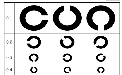
This is a familiar test using a mark that looks like a “C” of the alphabet. The size of the gap in the “C” helps determine visual acuity, such as 1.0 or 0.5. This mark is called a Landolt ring. Landolt is the name of a person, and the test method was devised by a French ophthalmologist named Landolt. |
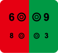 |
In addition, have you ever seen what is written on a red or green background? This is a test called the “Red-Green Test.” This test was conducted to confirm that the power of the spectacle lenses was neither too strong nor too weak. The “Red-Green Test” is based on a phenomenon called “chromatic aberration of the eye,” in which the position and size of the image differ depending on the wavelength (color) of light. The Red-Green Test is sometimes called the “Dichromatic Test.” |
2. Intraocular pressure test
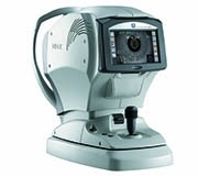 |
Intraocular pressure is like the air inside a ball if the eye is compared to a ball. It keeps the eyeball firm and helps maintain its size and shape. Intraocular pressure can be measured with a tonometer, a device that emits a puff of air toward the eye. Noncontact tonometers, which check the pressure by applying air to the eye’s surface, do not require anesthesia and are therefore commonly used for regular checkups and physical examinations. |
3. Visual field test
Stress and disease can cause a gradual loss of vision. It may seem that you would notice it when your vision begins to deteriorate, but it often goes unnoticed because it progresses slowly, your eyes become accustomed to it, and you can see things with both eyes. You can check your own visual field or have it tested by an ophthalmologist.
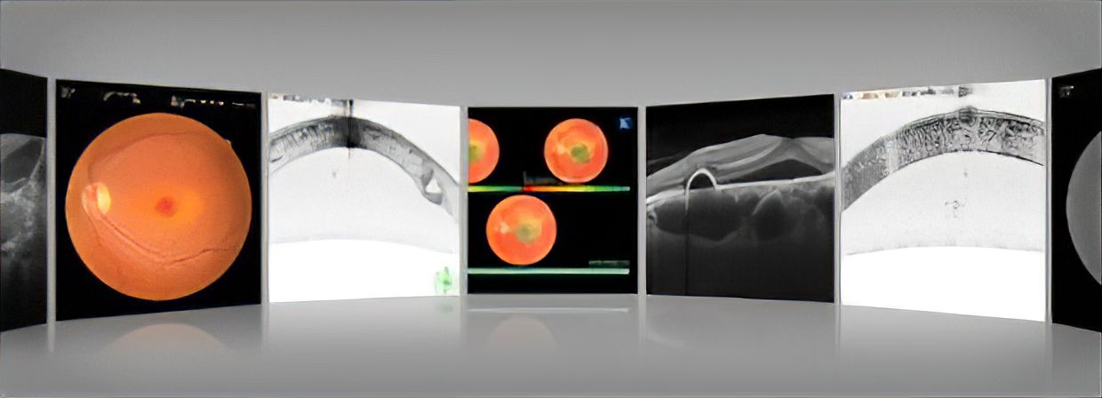
4. Fundus examination
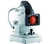 |
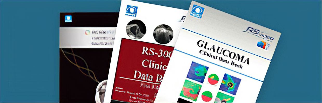
The bottom (back) of each eye was photographed with a device called a fundus camera. A camera can help diagnose retinal diseases, glaucoma, fundus hemorrhage, diabetes, arteriosclerosis, and hypertension by photographing and observing the back of the eye. |
Back to List