


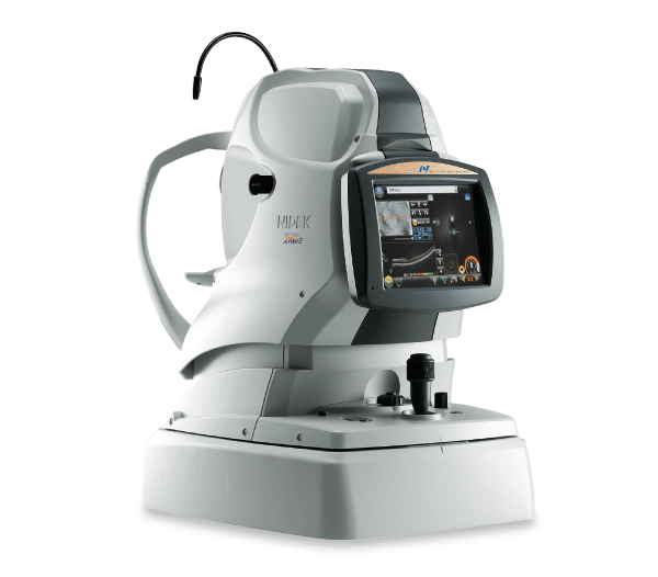
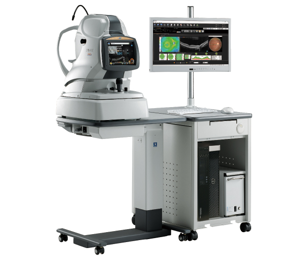
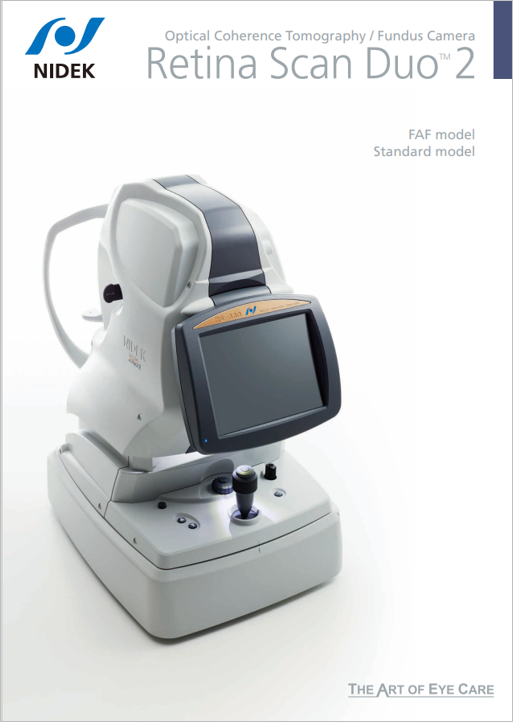
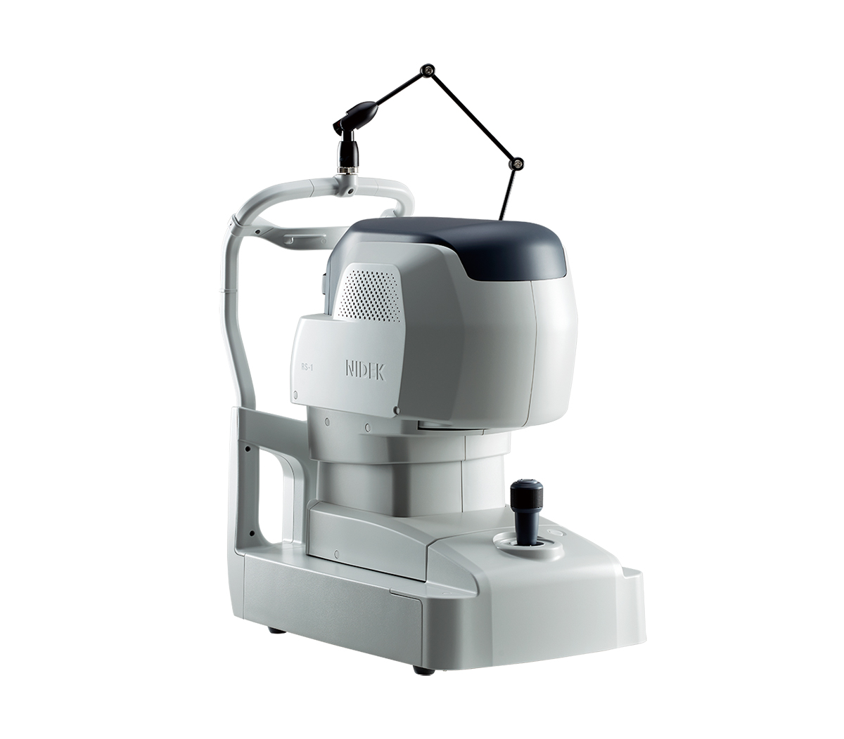
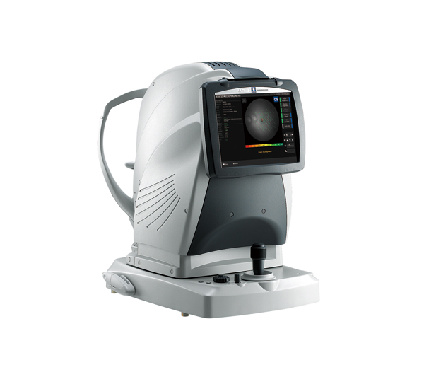
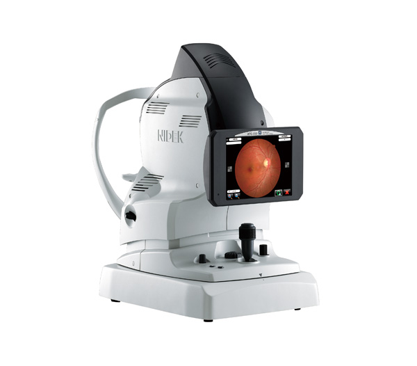
Optical Coherence Tomography / Fundus Camera
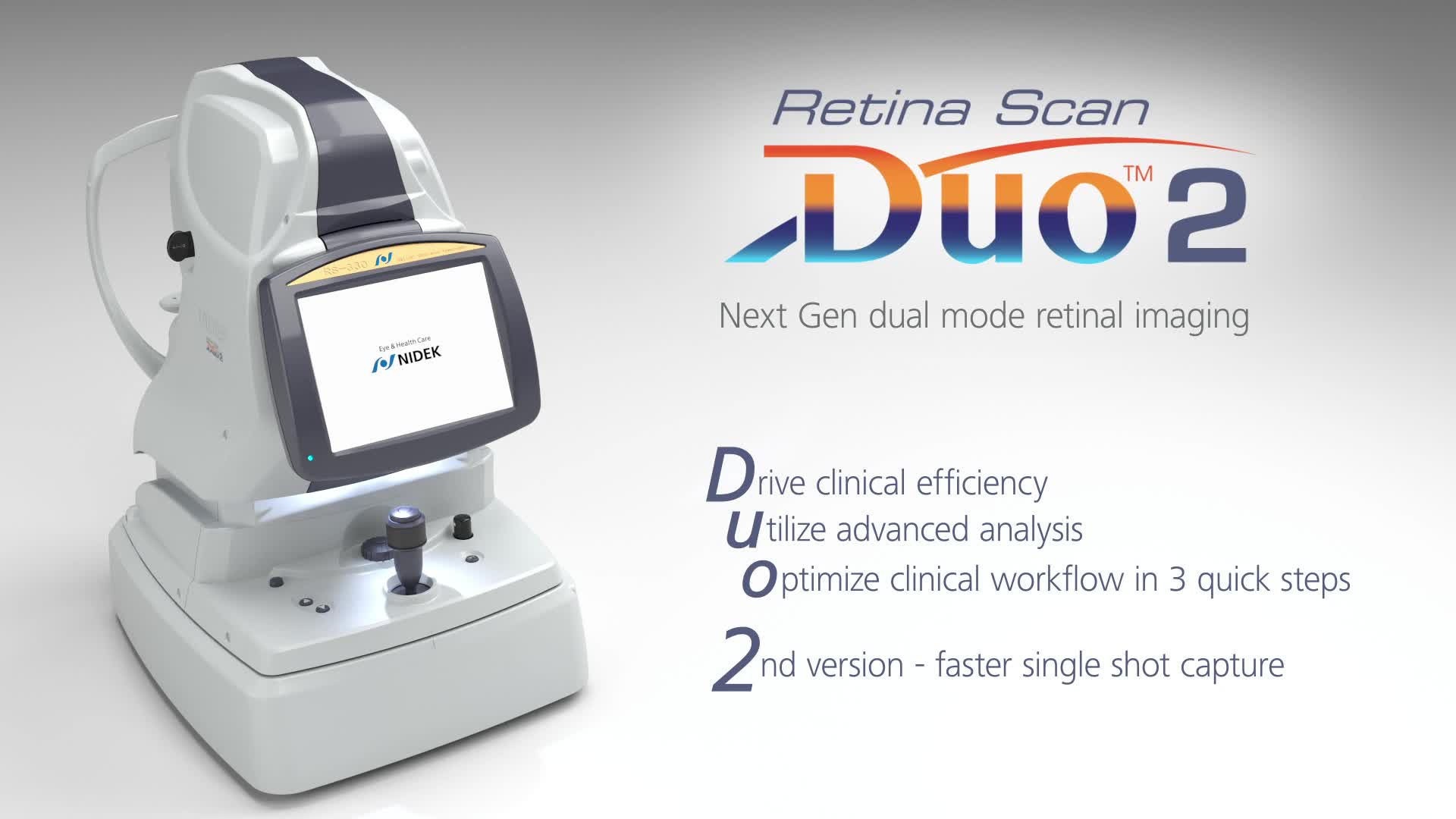
Retina Scan Duo™2
Features
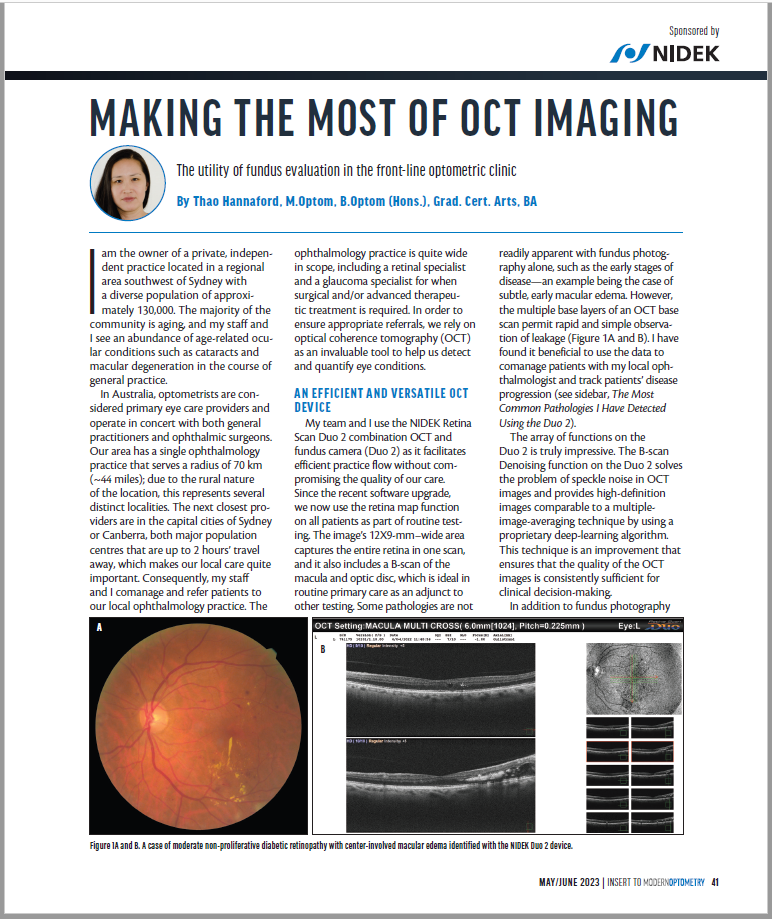
- Fundus image acquisition with macula and disc capture in one image on OCT
- Combined diagnosis of macular and disc pathologies
- Denoising technique with deep learning
- Quick acquisition of high definition B-scan images from a single-frame image
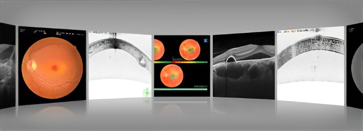
- Fundus autofluorescence (FAF)
Detailed Information
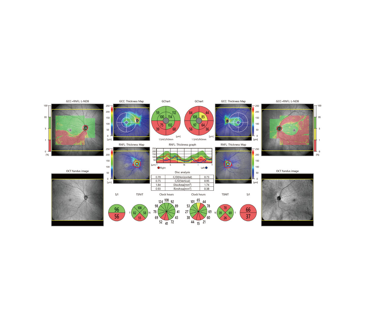
Combined diagnosis of macular and disc pathologies
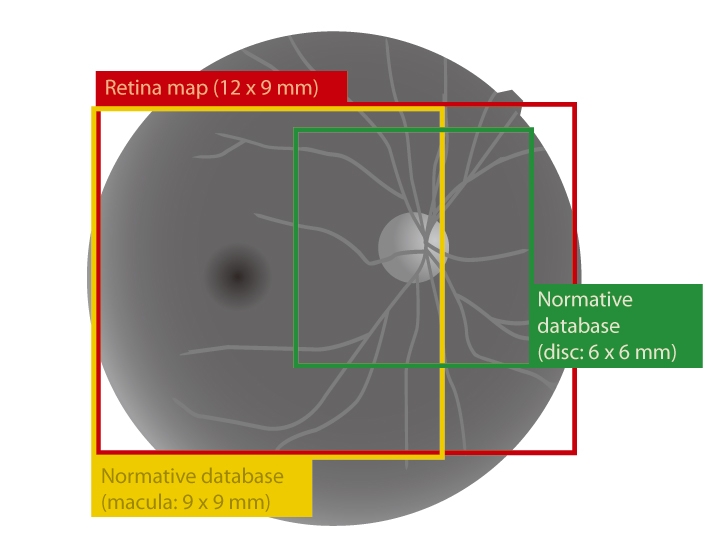
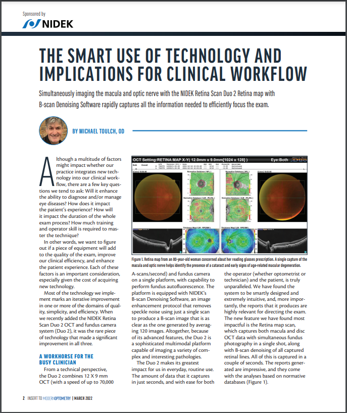
Retina map
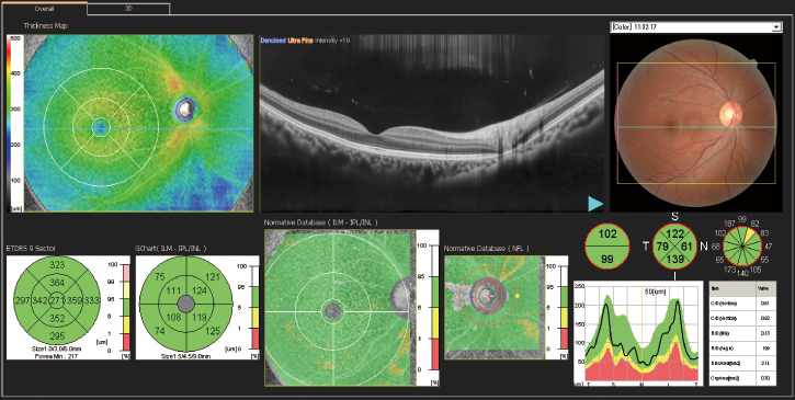
Wide area scan (12 x 9 mm)
Wide area normative database (macula: 9 x 9 mm, disc: 6 x 6 mm)
| A 12 x 9 mm wide area image can be acquired. The retina map captures both the macula and disc in a single shot.
The normative database provides a wide area color-coded map comparing the patient’s macular thickness to a population of normal eyes. |
 |
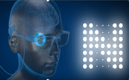
Denoising using deep learning
A new image enhancement technique using deep learning automatically displays a denoised image once B-scan acquisition is complete. With deep learning of a large data set of images averaged from 120 images, this denoising technique provides high definition images comparable to a multiple-image-averaging technique. The denoising function generates high definition images from a single frame while decreasing image acquisition time and increasing patient comfort.
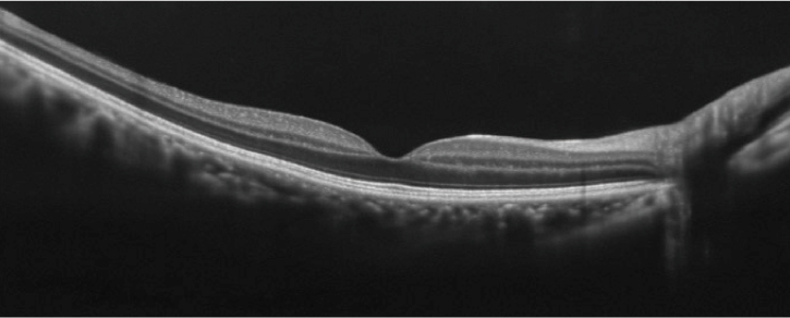
Denoised from a single-frame image |
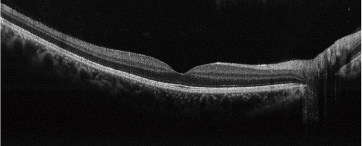
Averaged from 50 images |
Fundus autofluorescence (FAF)
| The FAF function is an advanced screening feature that allows non-invasive evaluation of the RPE without contrast dye.
*Available for the FAF model |
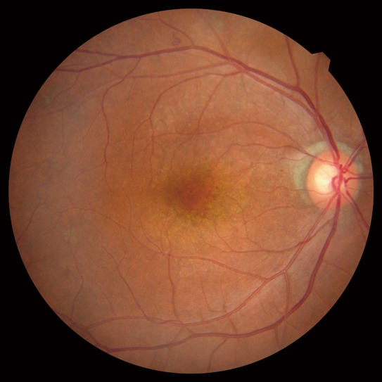
Color fundus image |
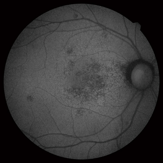
FAF image |
Optional features
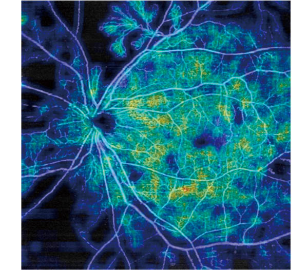
AngioScan
| Details are available on the AngioScan page. | 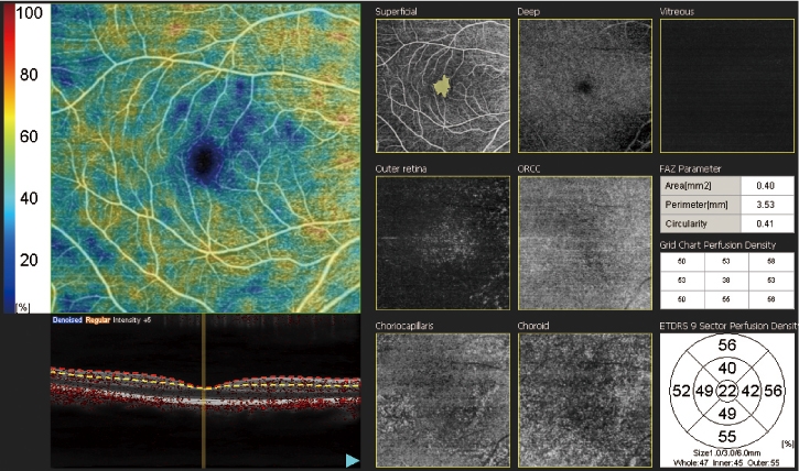 |
Long axial length normative database
Details are available on the long axial length normative database page.
Anterior segment adapter
The optional anterior segment adapter enables observation and analyses of the anterior segment.
Downloads
Videos
Product Information
|
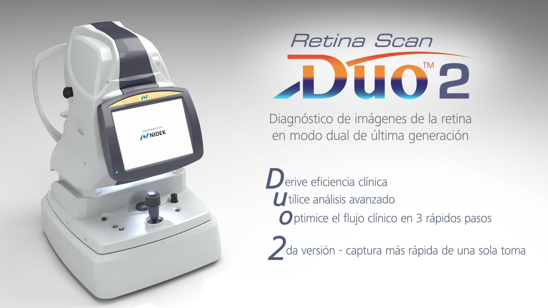
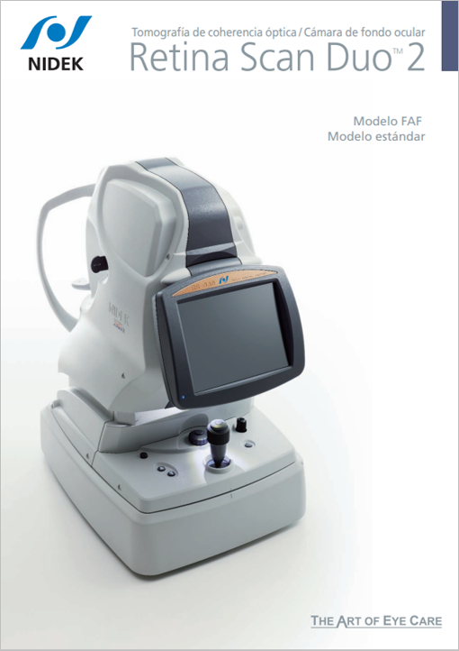
Introducción del producto (Spanish)
|
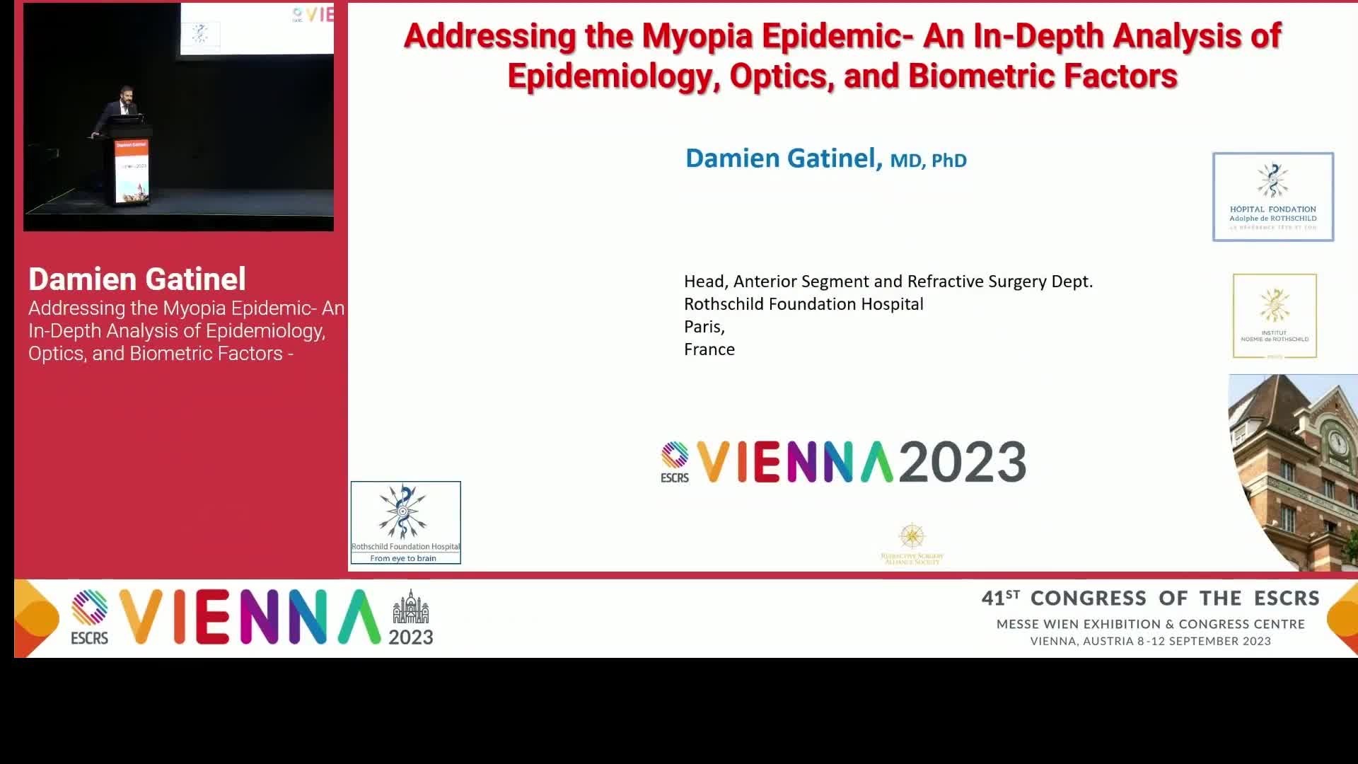
Lectures
|
User Testimonials
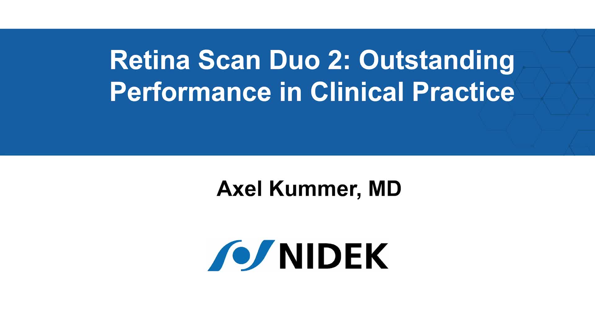
Axel Kummer, MD —Outstanding Performance in Clinical Practice
|
Related Products
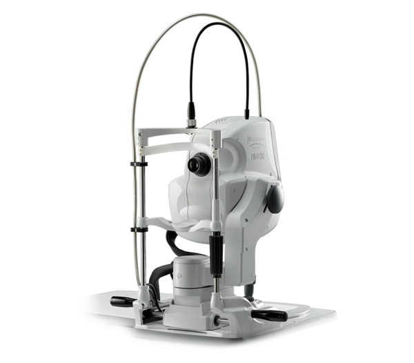
Scanning Laser Ophthalmoscope
Mirante SLO/OCT
Mirante SLO
Optical Coherence Tomography
RS-1 Glauvas
OCT-Angiography option for the Mirante SLO/OCT and Retina Scan Duo™ 2
Software for NIDEK OCT series
Long Axial Length Normative Database
Microperimeter
MP-3
Non-mydriatic Auto Fundus Camera
AFC-330
NOTE
The availability of products differs from country to country depending on the status of approval.
Specifications and design are subject to change without notice.
- Product/model name
- Optical Coherence Tomography RS-330