


Vol.20 The Formation and Function of the Eye—The Posterior Part of the Eye
| In a previous issue of Eye Story, Vol. 17, we introduced the “anterior eye” of the eye. This time, we will introduce the “posterior” part of the eye and its function. | 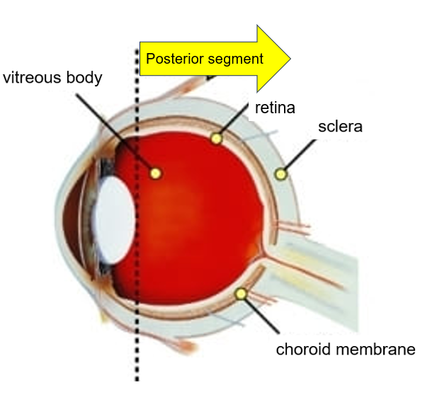 |
1. The vitreous body
The vitreous body is a colorless, transparent tissue that contains a large amount of water and looks jelly-like, similar to egg whites. Light passing through the lens travels through the vitreous body to the retina. The vitreous body maintains a constant distance between the lens and the retina, forming an appropriately sized image of the retina. The vitreous also acts as a cushion to prevent retinal damage.
2. Retina
The retina is a very thin membrane, approximately 0.2 mm thick, but is composed of 10 layers. It performs the most important function of seeing; if we compare it to a camera, it plays the role of a film. Light passing through the cornea, lens, and vitreous body forms an image that is transmitted as an electrical signal from the optic nerve to the brain.
3. Sclera
The sclera is an opaque white membrane approximately 1 mm thick that covers the white part of the eye and supports its structure.
4. Choroid
The choroid is a membrane containing numerous blood vessels inside the sclera. It is black and contains high concentration of melanin to prevent the reflection of incoming light. They also deliver oxygen and nutrients to the retina.
As you can see, the eye is a very complex organ. Although it is only 24 mm in diameter, it contains many tissues that work together to see objects.
Back to List