


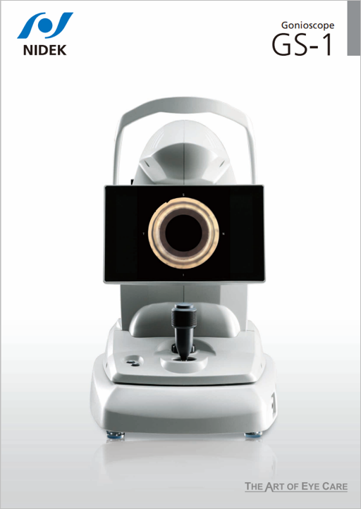
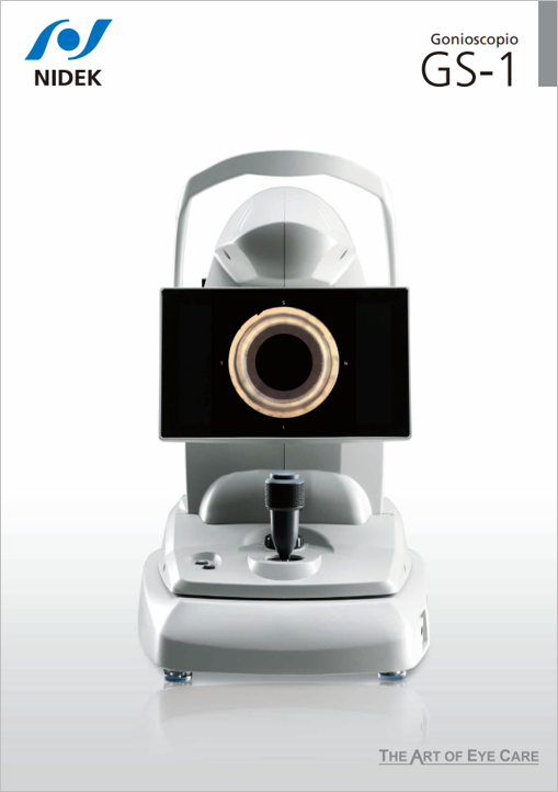
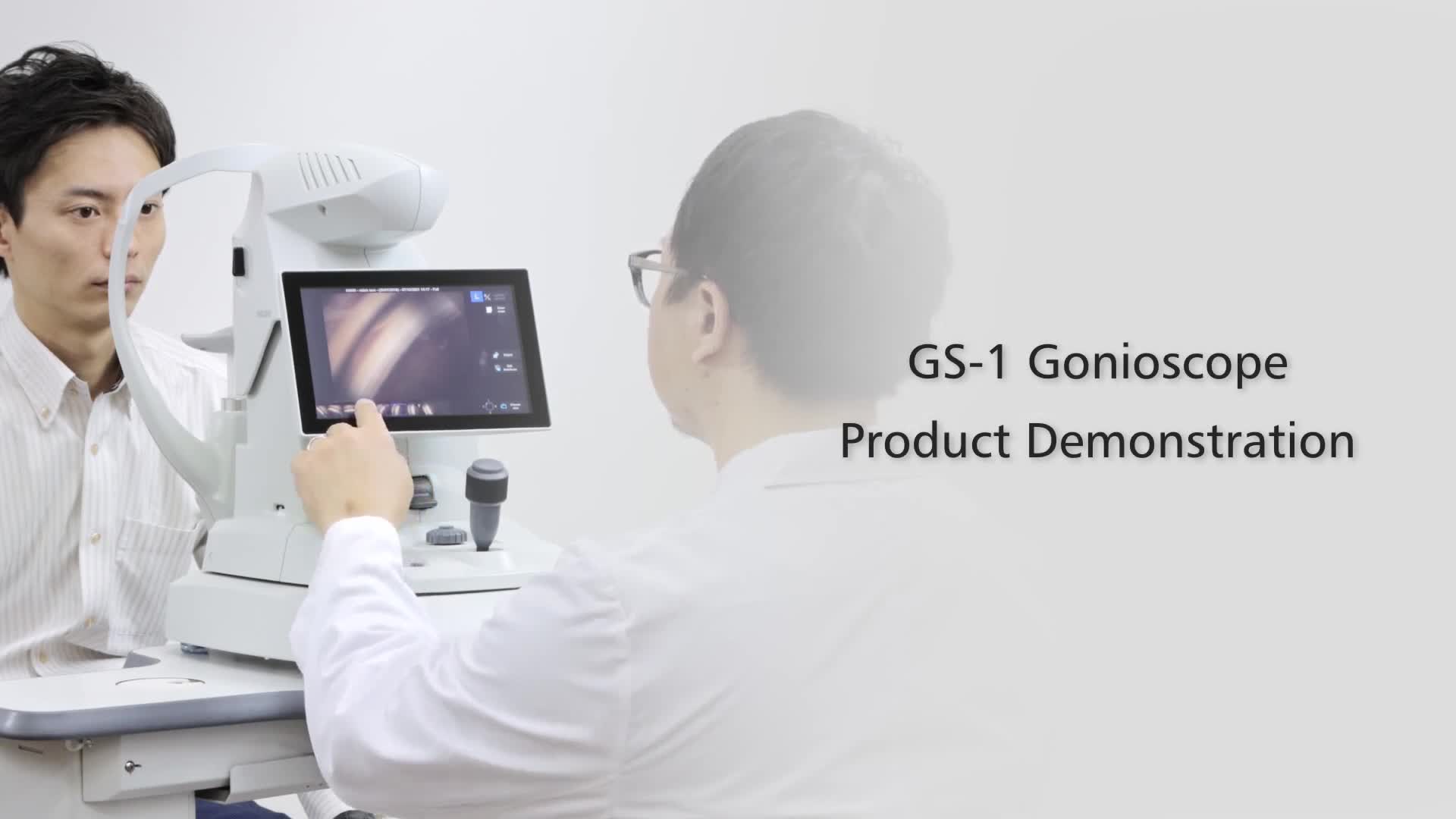
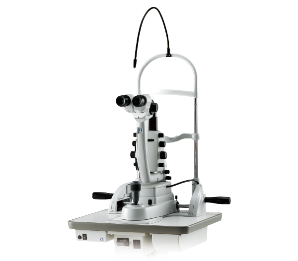
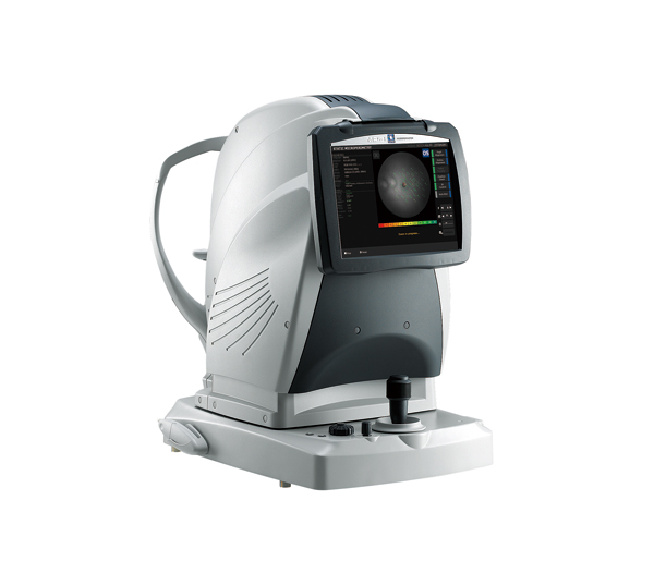
Gonioscope
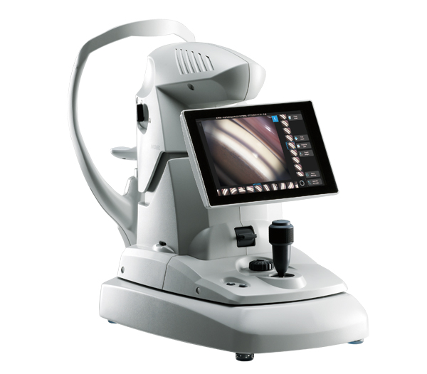
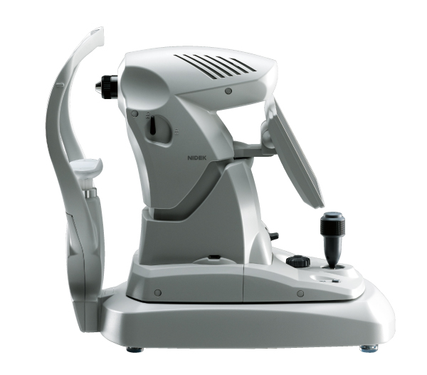
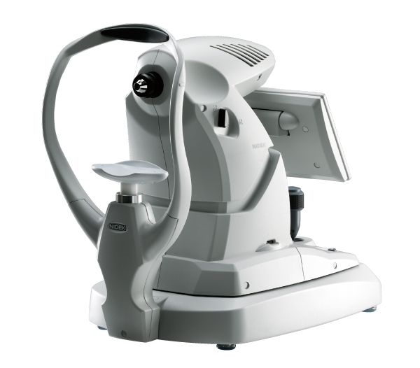
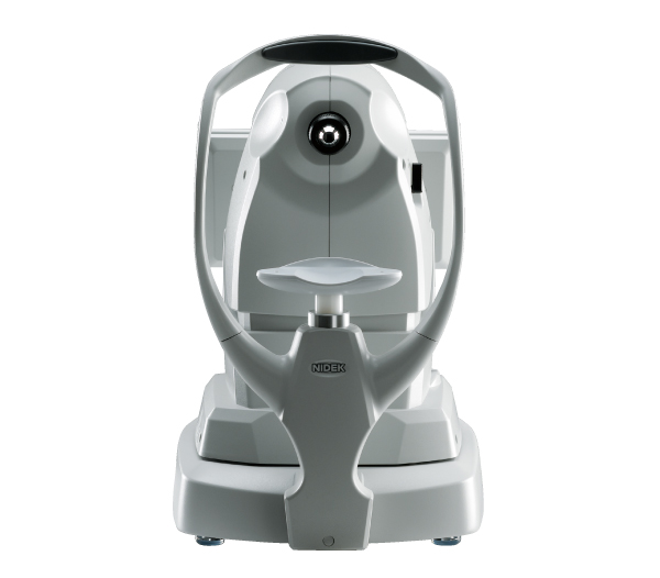
GS-1
Features
- Instant documentation of the iridocorneal angle in real-color photographs
- Freeing up time for the clinicians to assess and plan treatment
- Automated circumferential goniophotography
- Additional features with GS Viewer for NAVIS-EX
Detailed Information
Automated gonioscopy
Document. Assess. Plan.
A picture is worth a thousand words
 |
 |
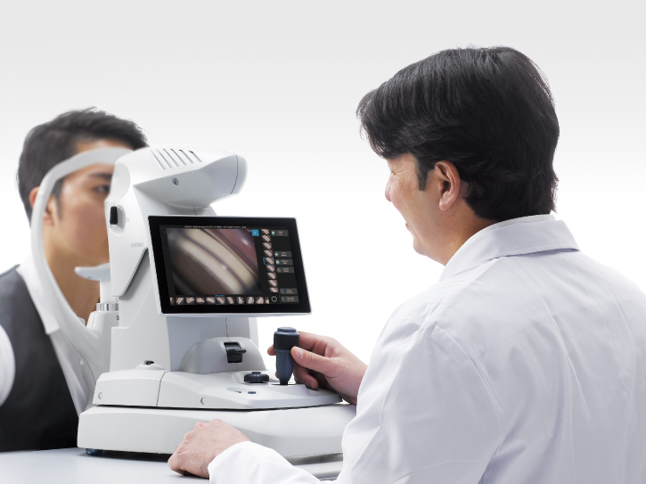
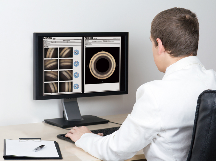
Instant documentation with the GS-1
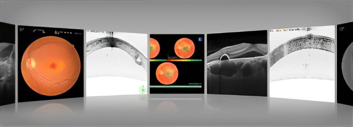
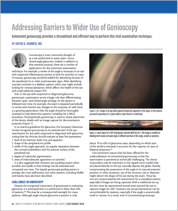
The GS-1 instantly documents the iridocorneal angle in real-color photographs and stores them in the device. Hence, there is no need for detailed drawing or sketches in the patient chart to record angle pathology. Photographs from the GS-1 can be attached to the patient file or chart. Documentation with the GS-1 is far simpler and much more definitive than subjective sketches of pathology.
Easy documentation and assessment with the GS-1. |
The photographs can be checked on the touch screen of the device and easily magnified. |
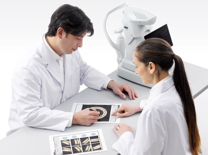
Documentation to be shared
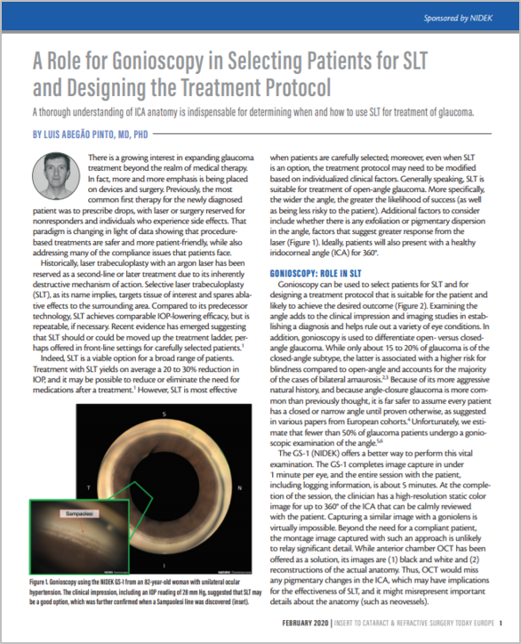
Assessment of angle structure and pathology is easily facilitated by reviewing the digital photographs during clinical evaluation rather than performing manual gonioscopy. The digital photographs from the GS-1 can be shared from technicians to clinicians, with fellow clinicians, the referring physicians and patients, alongside complete documentation of GS-1 findings.

Focus on assessment and planning
|

Consultation with fellow clinicians
|
Effective information transfer to partner clinics |

Easier patient consent |
Assess and plan with documentation
The GS-1 frees up time for the clinicians to assess and plan treatment. The digital goniophotographs add the convenience of re-assessing the entire angle at any time. High resolution color photographs enhance the quality of assessment and allow comprehensive follow-up.
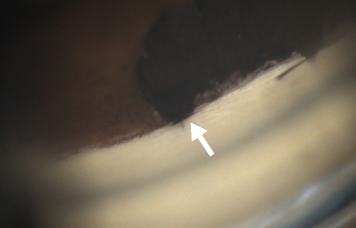
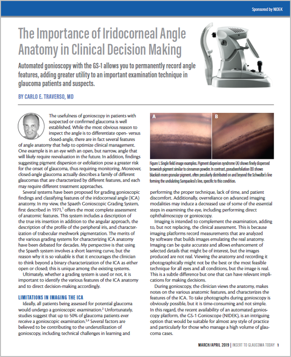
Postoperative examples*
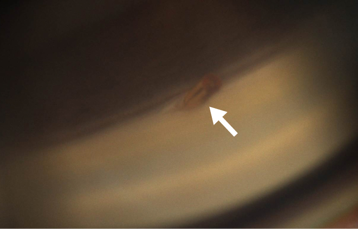
MIGS
|
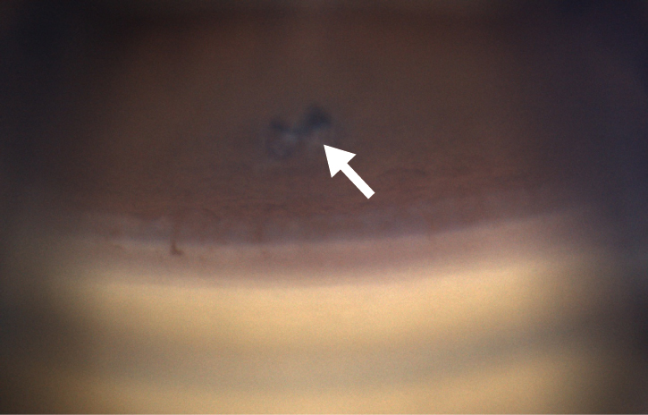
Laser iridotomy
|

Tube |
Trabeculectomy |
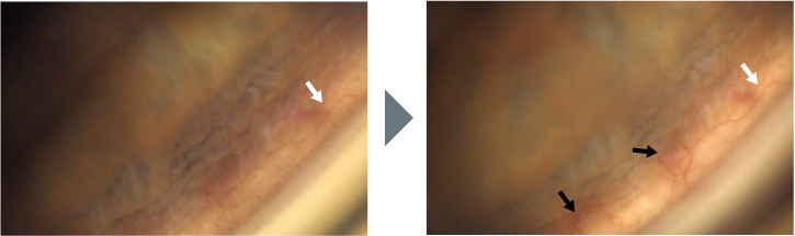
Progression of neovascularization (after 5 months)*
* Image courtesy of Prof C. E. TRAVERSO, MD, Clinica Oculistica, Di.N.O.G.M.I., University of Genova – Ospedale Policlinico S. Martino, Italy
GS-1 features
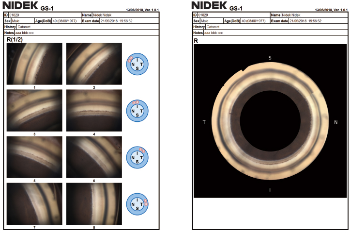
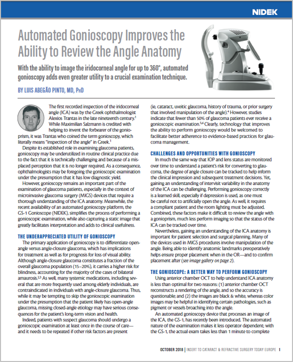
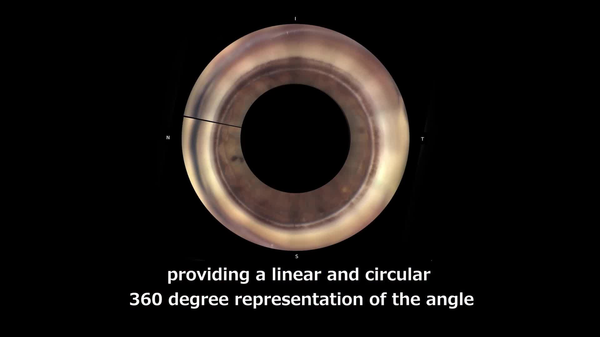
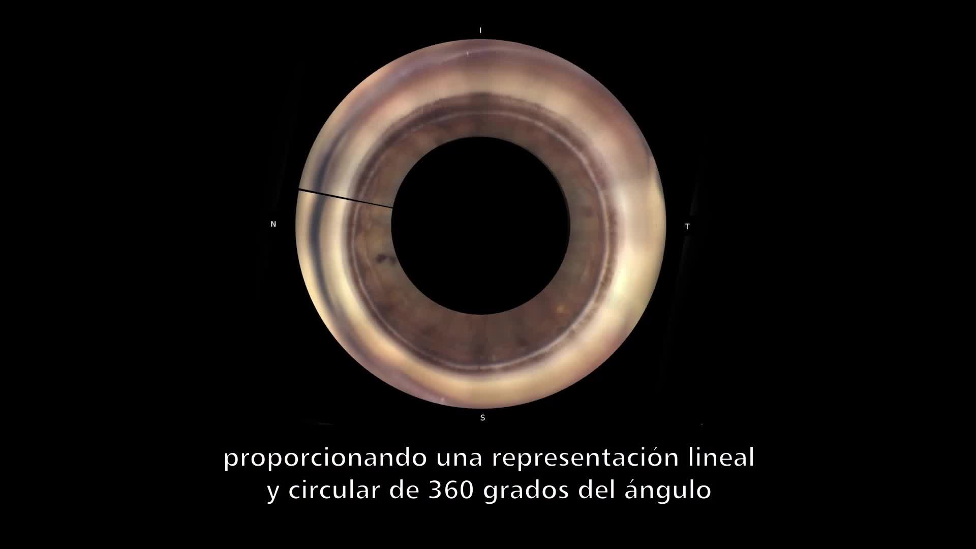
Automated circumferential goniophotography
| An internal optical system automatically rotates and acquires color photographs of the iridocorneal angle in 16 directions / 360 degrees documenting the entire angle. Each direction can be captured in 17 different foci, enabling a versatile approach to iridocorneal angle photography. | 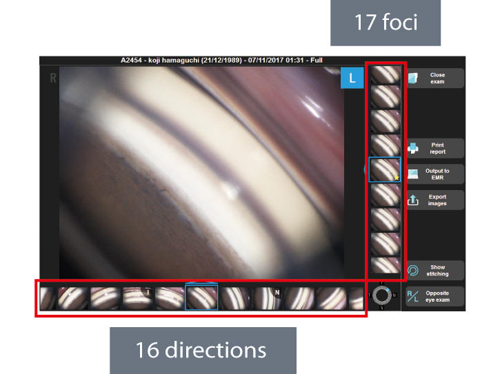 |
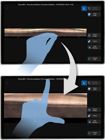
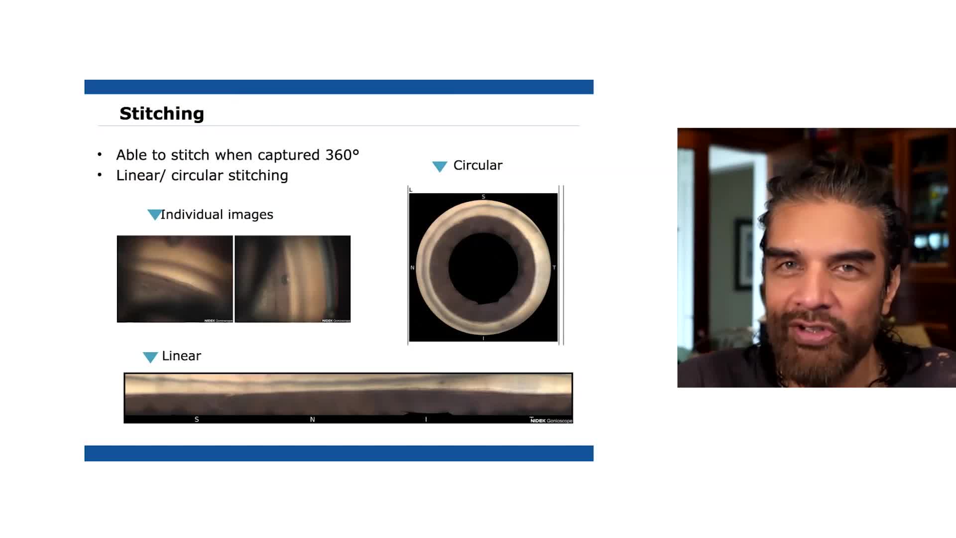
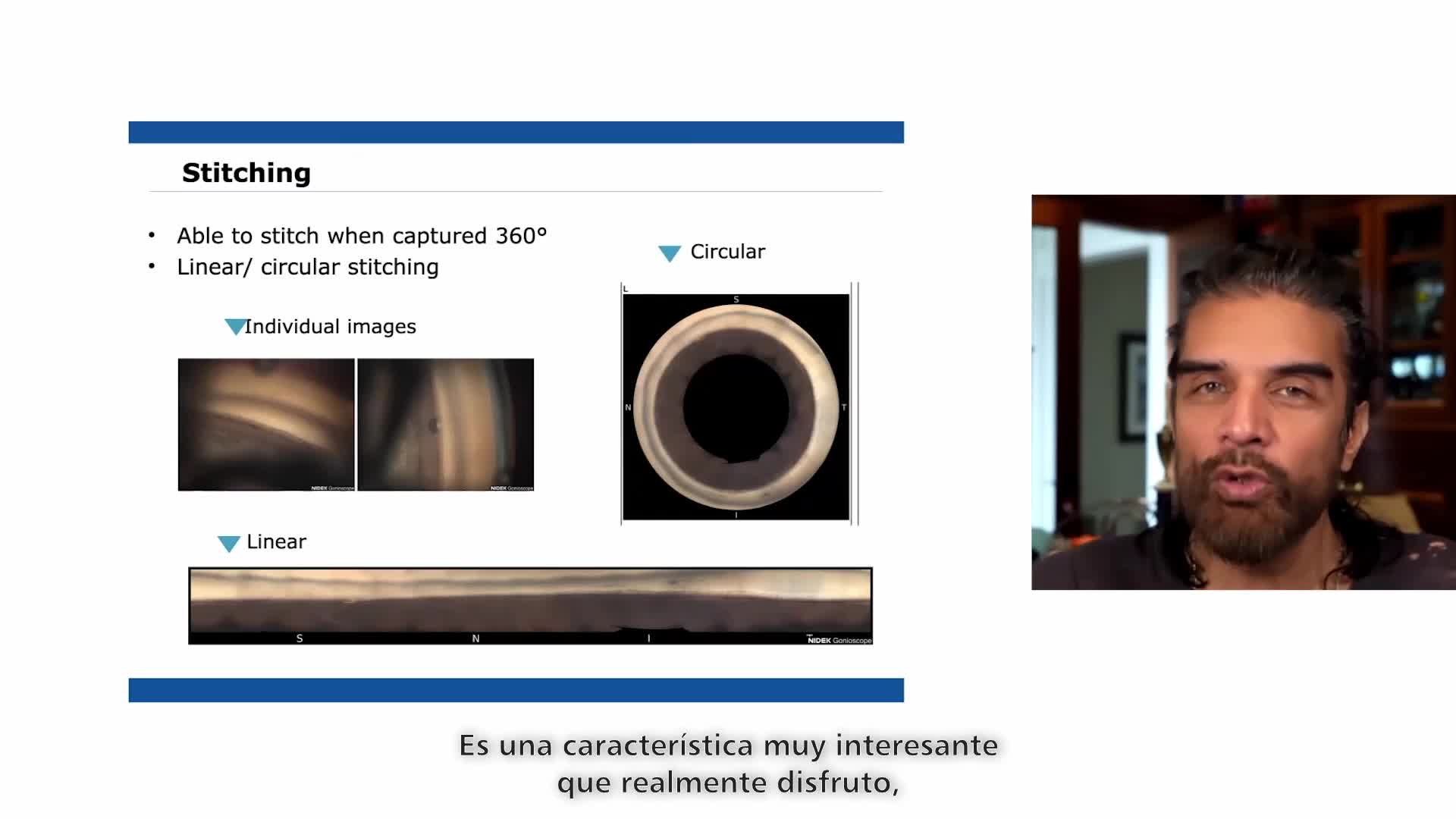
Export images
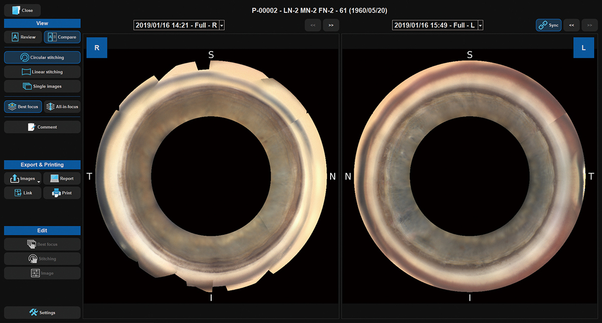
| The images acquired by the GS-1 can be displayed as a single image, with circular stitching, or linear stitching. In addition to detailed assessment with single image, stitching allows localization of features/pathologies within the entire angle. High-resolution color images are exported in JPEG, PNG and PDF files. | 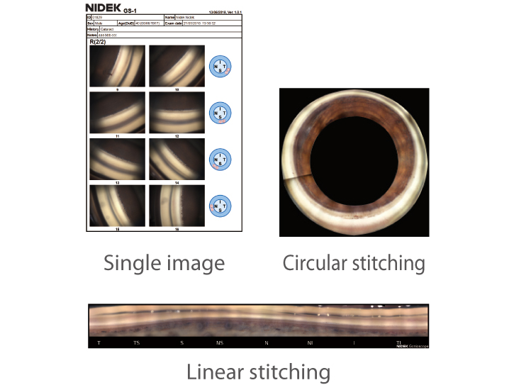 |
Non-contact gel immersion measurement
| To ensure patient comfort, a coupling gel is used during image acquisition. The multimirror prism is not intended to touch the cornea. | 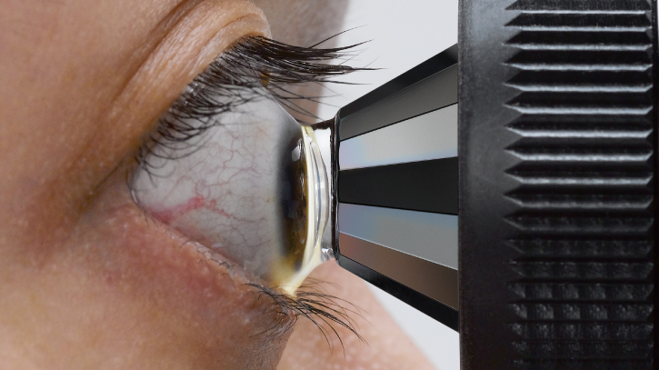 |
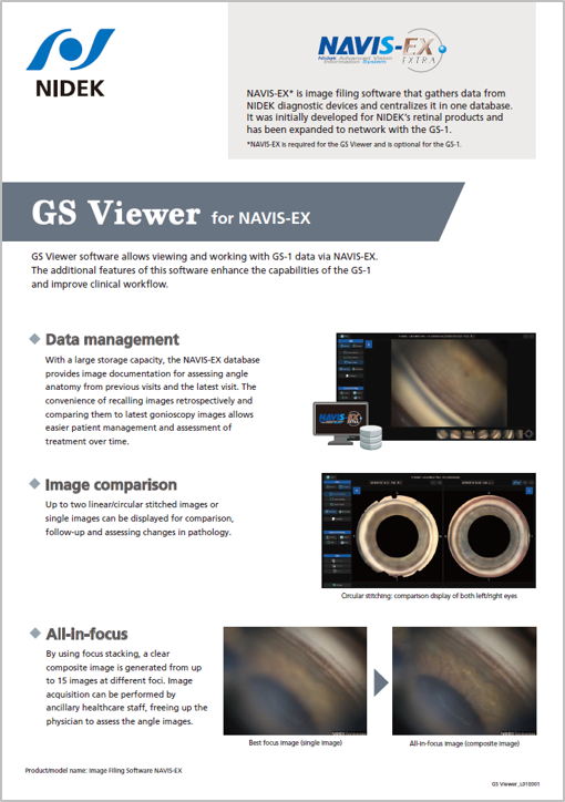
Additional features with GS Viewer for NAVIS-EX* (optional)
GS Viewer software allows viewing and working with GS-1 data via NAVIS-EX. The additional features of this software enhance the capabilities of the GS-1 and improve clinical workflow.
*NAVIS-EX is required for the GS Viewer and is optional for the GS-1. Product/model name: Image Filing Software NAVIS-EX
Data management
| With a large storage capacity, the NAVIS-EX database provides image documentation for assessing angle anatomy from previous visits and the latest visit. The convenience of recalling images retrospectively and comparing them to latest gonioscopy images allows easier patient management and assessment of treatment over time.
|
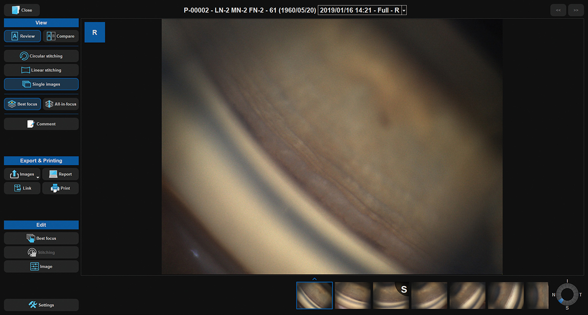 |
Image comparison
| Up to two linear/circular stitched images or single images can be displayed for comparison, follow-up and assessing changes in pathology.
|
|
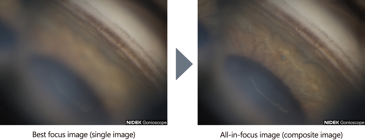
All-in-focus
| By using focus stacking, a clear composite image is generated from up to 15 images at different foci. Image acquisition can be performed by ancillary healthcare staff, freeing up the physician to assess the angle images.
|
 |
|
Downloads

Videos
User Testimonials

Carlo E. Traverso, MD
Professor and Chairman, Clinica Oculistica University of Genova.
Policlinico Ospedale San Martino IRCCS, Italy

Fernando Gomez Goyeneche, MD
Emeritus Professor Hospital Militar Central, Colombia

Luis Abegão Pinto, MD, PhD
Assistant Professor, Lisbon University, Portugal

Alvaro Dantas, MD
ICONE- Instituto de Cirurgia Ocular do Nordeste (Northeast Eye Surgery Institute), Brazil

Carlo Alberto Cutolo, MD, PhD
Clinica Oculistica University of Genova.
Policlinico Ospedale San Martino IRCCS, Italy
Carlo E. Traverso, MD
Professor and Chairman, Clinica Oculistica University of Genova.
Policlinico Ospedale San Martino IRCCS, Italy
Fernando Gomez Goyeneche, MD
Emeritus Professor Hospital Militar Central, Colombia
Luis Abegão Pinto, MD, PhD
Assistant Professor, Lisbon University, Portugal
Alvaro Dantas, MD
ICONE- Instituto de Cirurgia Ocular do Nordeste (Northeast Eye Surgery Institute), Brazil
Carlo Alberto Cutolo, MD, PhD
Clinica Oculistica University of Genova.
Policlinico Ospedale San Martino IRCCS, Italy
Related Products
Slit Lamp
SL-2000
Optical Coherence Tomography
RS-1 Glauvas
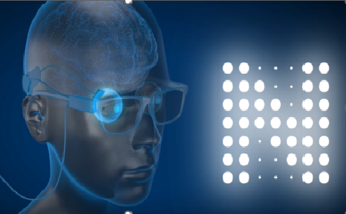
Microperimeter
MP-3
Ophthalmic YAG and SLT Laser System
YC-200 S plus
Ophthalmic YAG Laser System
YC-200
Specular Microscope
CEM-530
NOTE
The availability of products differs from country to country depending on the status of approval.
Specifications and design are subject to change without notice.
- Product/model name
- GONIOSCOPE GS-1