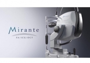
Room Mirante
Use the Center Map to explore your interest in the Mirante.
Center Map
Main Auditorium
Watch sessions on demand
Lecture Hall
What do doctors say about Mirante?
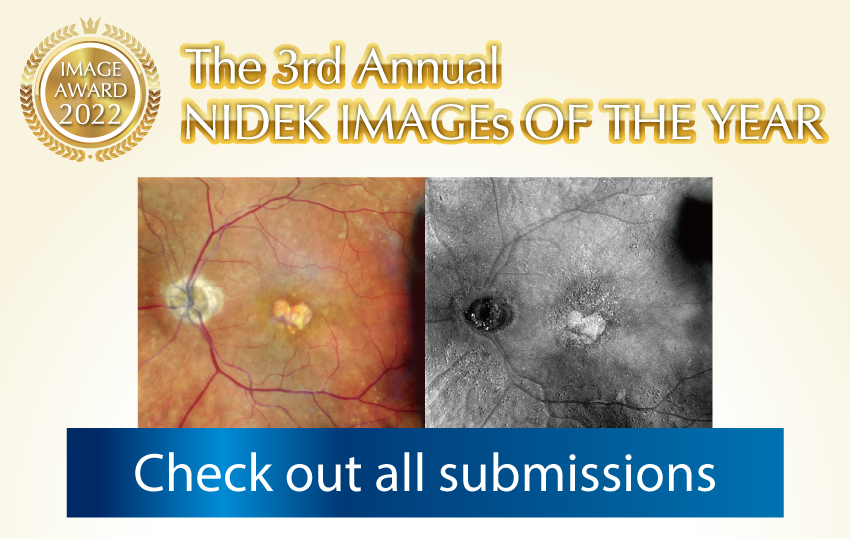
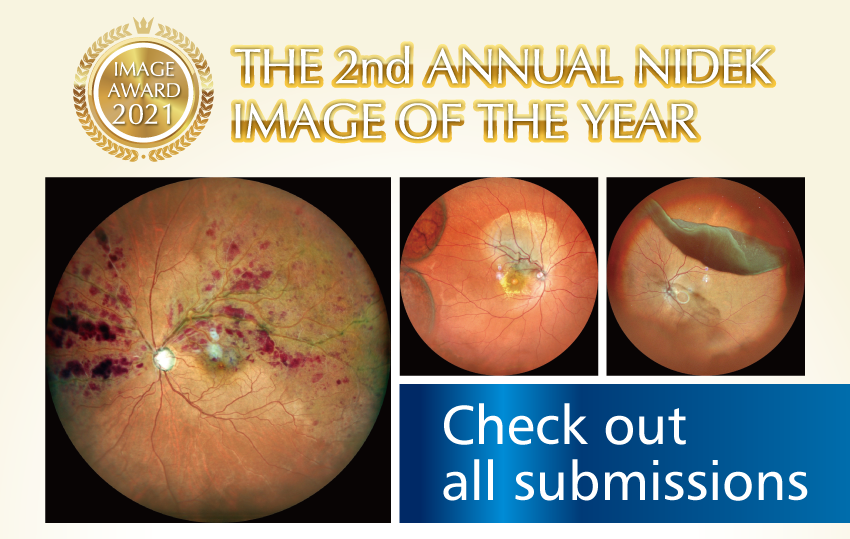
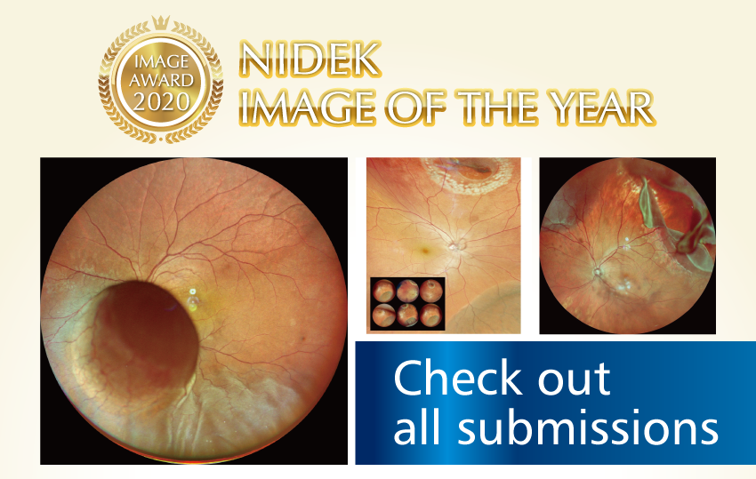
Image Gallery
Clinically stunning images from your peers
Room 101
Basics of the Mirante including operation
Room 201
“Must know” functions of Mirante
Learning Lounge
Technical expertise and more
Some exclusive content is available only to NIDEK web members.
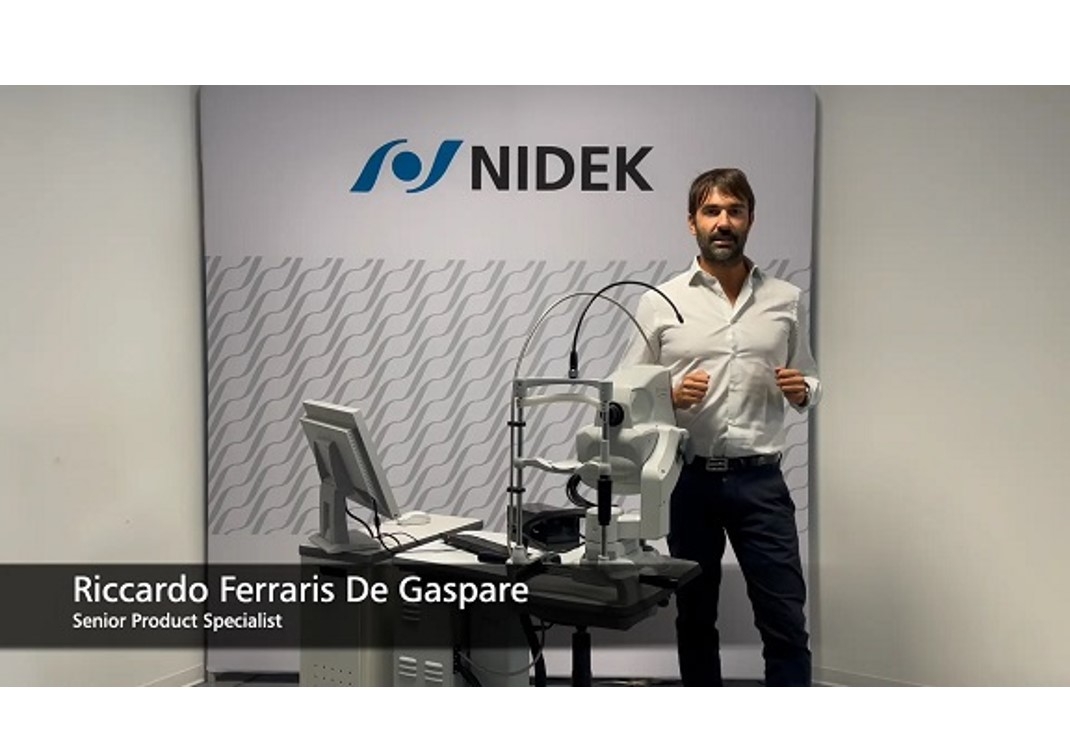
Mirante testimonials – Why I use Mirante

Main Auditorium – Watch sessions on demand
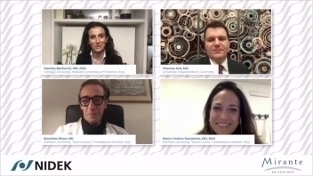
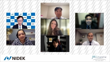
Lecture Hall – What do doctors say about Mirante?
Video
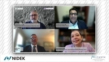

Utilizing Multimodal & Ultra Wide Field Imaging in the Diagnosis and Management of Retinal Diseases
SriniVas R. Sadda, MD, K. Bailey Freund, MD, Nadia Waheed, MD
Reading Time
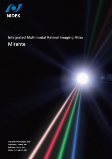
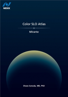
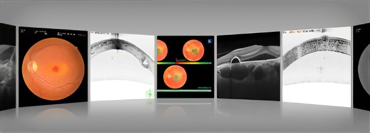
Image Gallery – Clinically stunning images from your peers
Room 101 – Basics of the Mirante including operation
▲Back to Center MapRoom 201 – “Must know” functions of Mirante
Retro mode bibliography
- Cozzi M, Monteduro D, Parrulli S, et al. Sensitivity and Specificity of Multimodal Imaging in Characterizing Drusen. Ophthalmol Retina. 2020;4(10):987-995. doi:10.1016/j.oret.2020.04.013
- Corradetti G, Byon I, Corvi F, Cozzi M, Staurenghi G, Sadda SR. Retro mode illumination for detecting and quantifying the area of geographic atrophy in non-neovascular age-related macular degeneration. Eye (Lond). 2022;36(8):1560-1566. doi:10.1038/s41433-021-01670-3
- Savastano A, Ripa M, Savastano MC, et al. Retromode Imaging Modality of Epiretinal Membranes. J Clin Med. 2022;11(14):3936. Published 2022 Jul 6. doi:10.3390/jcm11143936
- Savastano MC, Kilian RA, Savastano A, et al. Morphological Features of Full-Thickness Macular Holes Using Retromode Scanning Laser Ophthalmoscopy. Ophthalmic Surg Lasers Imaging Retina. 2022;53(7):368-373. doi:10.3928/23258160-20220614-01
- Corradetti G, Corvi F, Sadda SR. Subretinal Drusenoid Deposits Revealed by Color SLO and Retro-Mode Imaging. Ophthalmology. 2021;128(3):409. doi:10.1016/j.ophtha.2020.09.038
- Mainster MA, Desmettre T, Querques G, Turner PL, Ledesma-Gil G. Scanning laser ophthalmoscopy retroillumination: applications and illusions. Int J Retina Vitreous. 2022;8(1):71. Published 2022 Sep 30. doi:10.1186/s40942-022-00421-0










 TOP
TOP


