


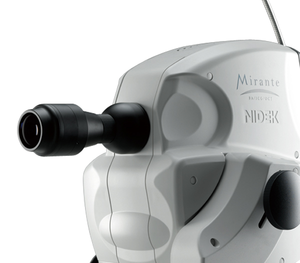
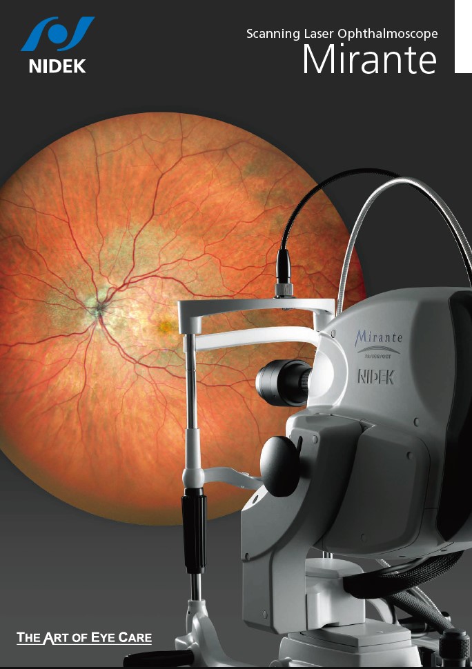
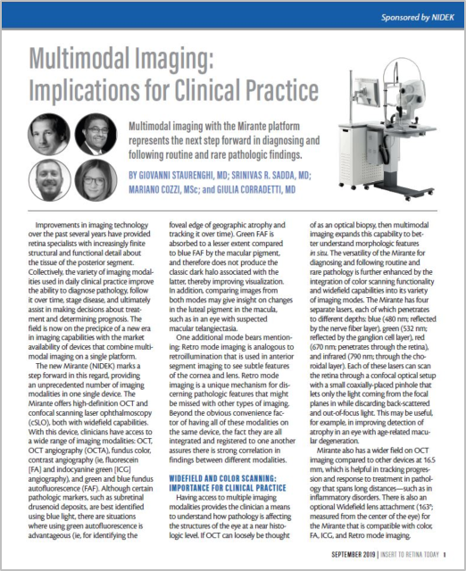
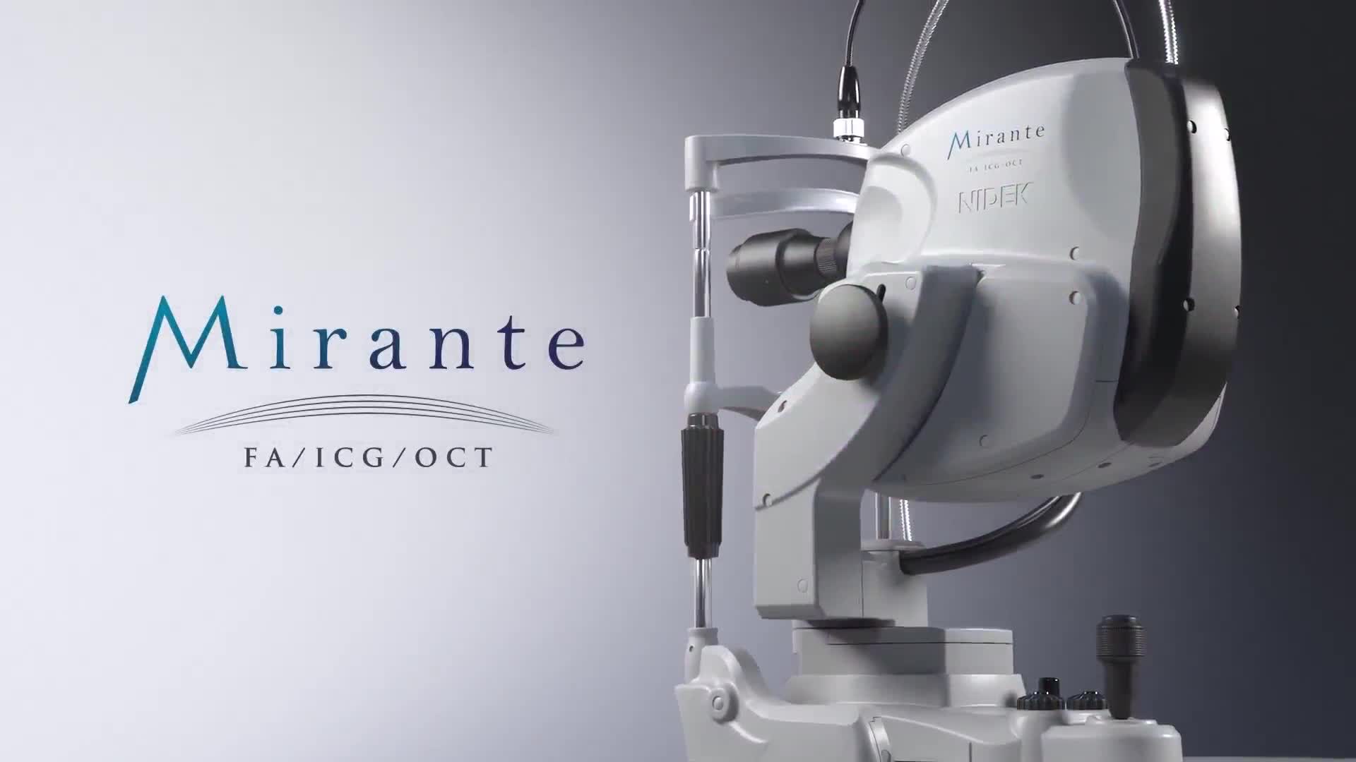
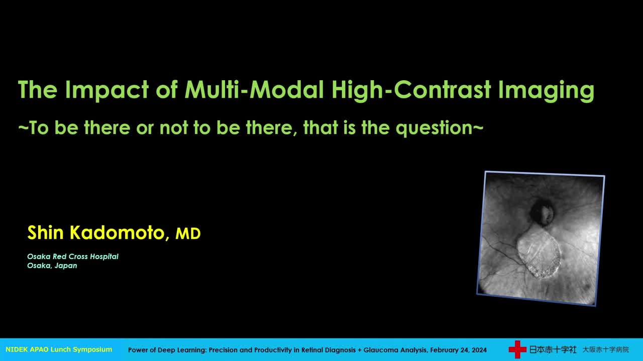
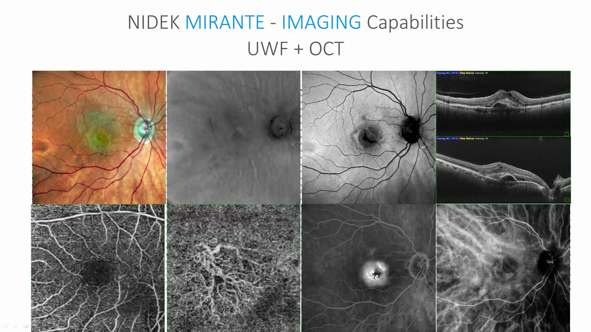
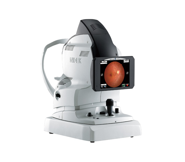
Scanning Laser Ophthalmoscope
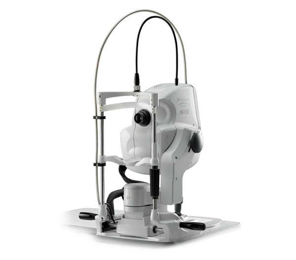
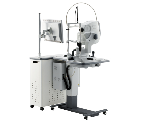
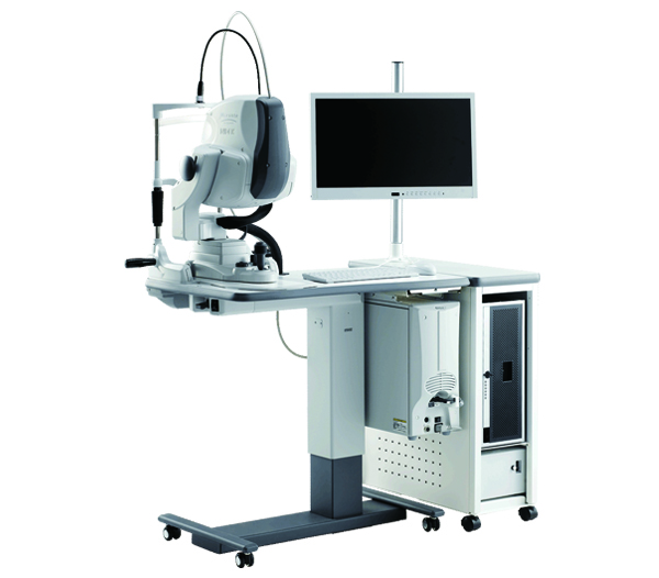
Mirante SLO/OCT
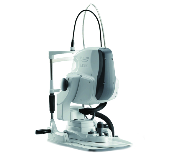
Mirante SLO
Features
- The ultimate multimodal imaging platform
For the SLO/OCT model
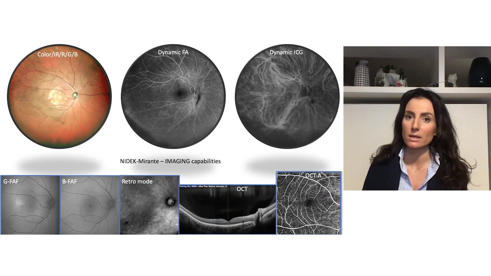
– Color / FA / ICG / Blue-FAF / Green-FAF / Retro mode
– OCT / OCT-Angiography*
For the SLO model
– Color / FA* / ICG* / Blue-FAF / Green-FAF / Retro mode - Ultra wide field x ultra HD image*
- Unsurpassed color
- Dynamic/Simultaneous FA and ICG
- Unique Retro mode
- HD wide area OCT
- Fly Through function*Optional
Detailed Information
The ultimate multimodal imaging platform
For the SLO/OCT model
– Color / FA / ICG / Blue-FAF / Green-FAF / Retro mode
– OCT / OCT-Angiography*
For the SLO model
– Color / FA* / ICG* / Blue-FAF / Green-FAF / Retro mode
*Optional
 |
 |
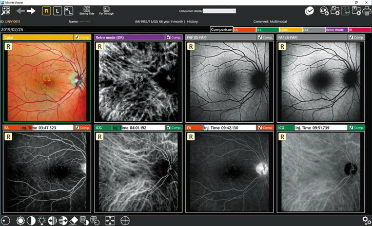
Streamlined combination capture
The Combo image capture allows sequential capture of images with the preset combination of image capture settings for each specified disease.
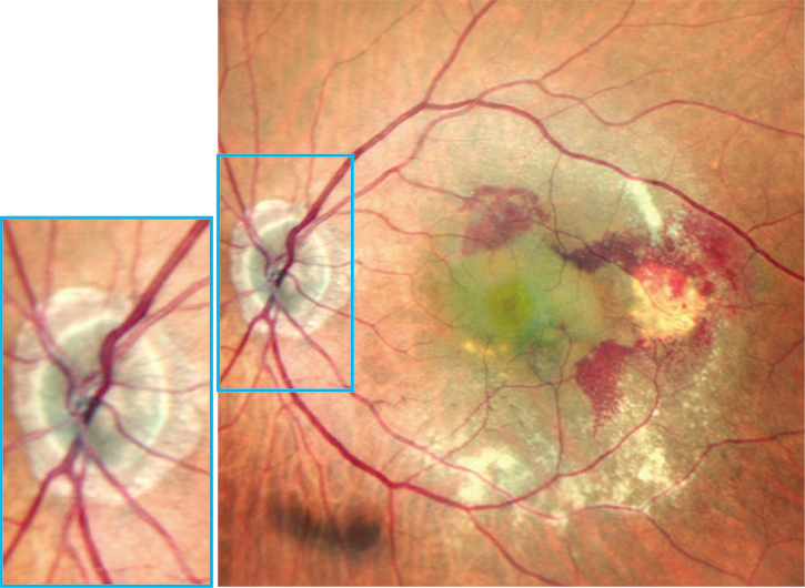
Ultra wide field x ultra HD image*
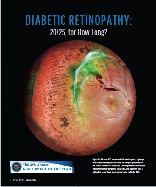
163° ultra wide field image
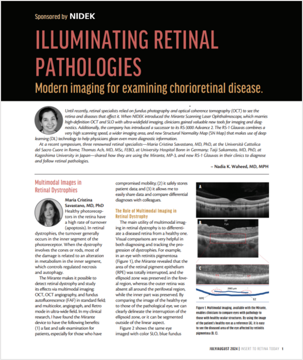
The clear image of the entire 163° field of view** enables detailed evaluation of pathologies from the fovea to the extreme periphery.
* Ultra wide field imaging is available with the optional wide-field adapter.
** Measured from the center of the eye
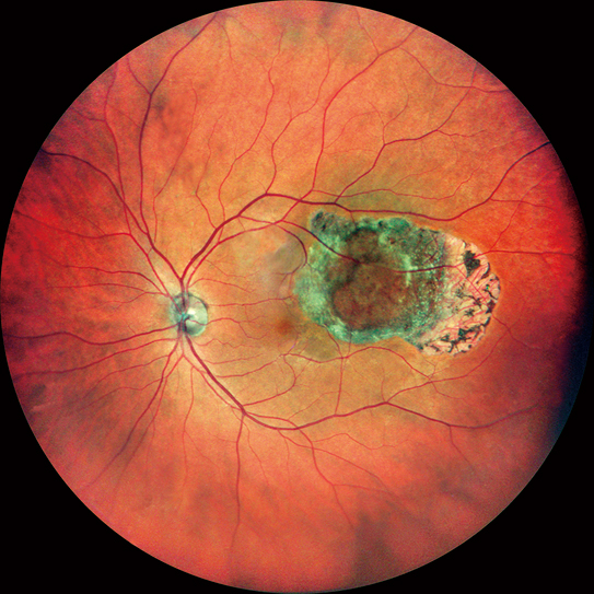
Color
|
Retro mode
|
FA |
ICG |
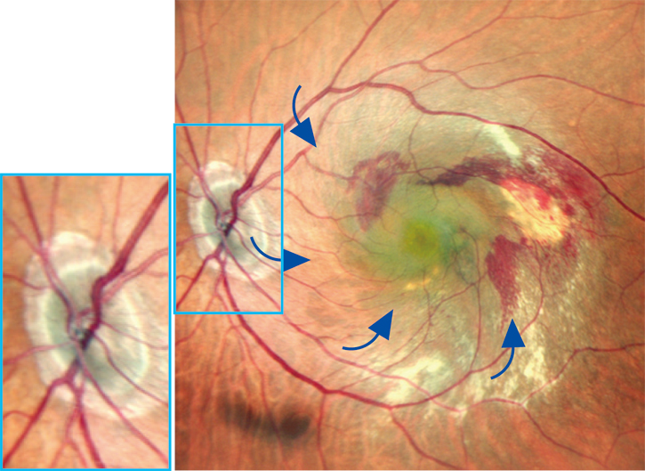
Ultra 4K HD and averaging function for unparalleled clarity
4,096 x 4,096 pixel imaging captures every detail of the retina and choroid. Additionally, zooming in allows high magnification, clear visualization of subtle changes in pathology, and resolution of the fine details of capillaries.
The FlexTrack algorithm corrects image distortion due to unstable fixation and enhances averaging quality.
Distorted image due to poor fixation |
Corrected image using FlexTrack |
Unsurpassed color
Three separate RGB detectors simultaneously scan different depths of retina with red, green, and blue wavelengths. A color histogram is available for fine adjustment based on pathology or practitioner preference.
Color histogram adjusted similar to slit lamp view |
Color histogram adjusted similar to fundus camera image |
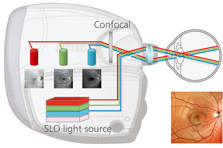
RGB detectors (Light-sensitive elements) |
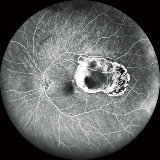
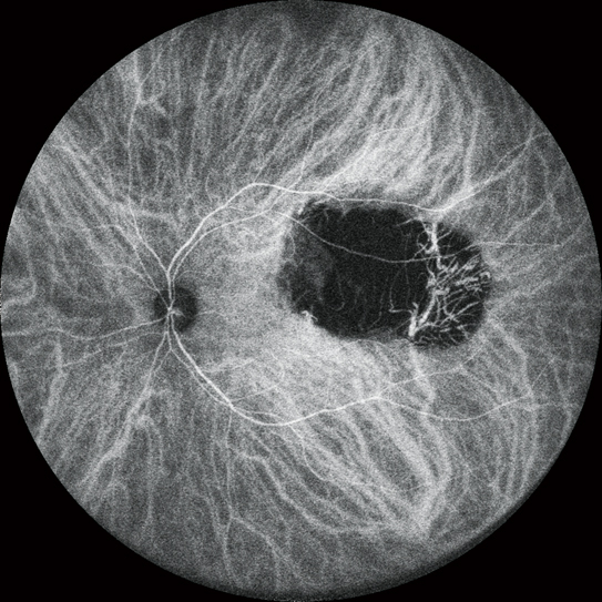
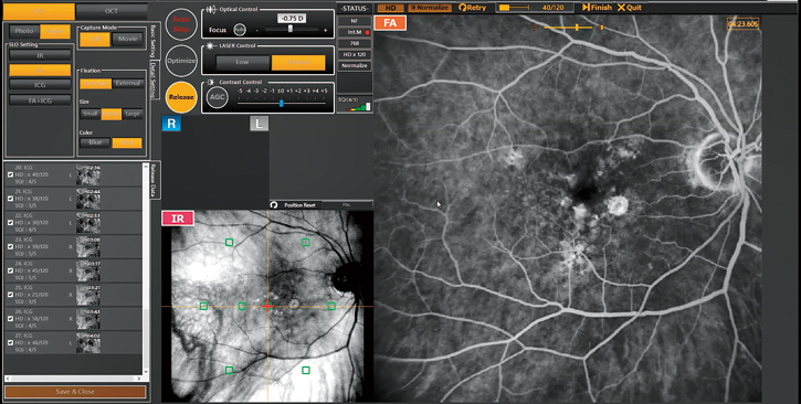
FA and ICG*
HD dynamic angiogram
| Videos can be recorded at a maximum of 1,024 x 1,024 pixels for up to 120 seconds. Multiple short videos can be recorded during the same measurement. | 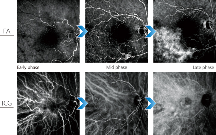 |
Simultaneous FA and ICG
The Mirante allows simple, simultaneous acquisition of FA and ICG images.
The live IR monitoring enables alignment prior to fluorescence emission and reduces the risk of missing the very early phase of angiography.
The Auto gain control (AGC) simultaneously adjusts contrast of each FA and ICG image, making the imaging of dynamic blood flow a very simple procedure.
*Available for the SLO/OCT model. Optional for the SLO model.
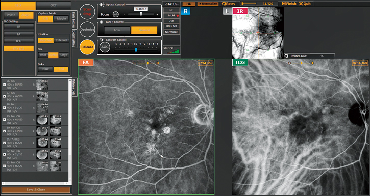
Simultaneous FA and ICG imaging display |
Live IR monitoring |
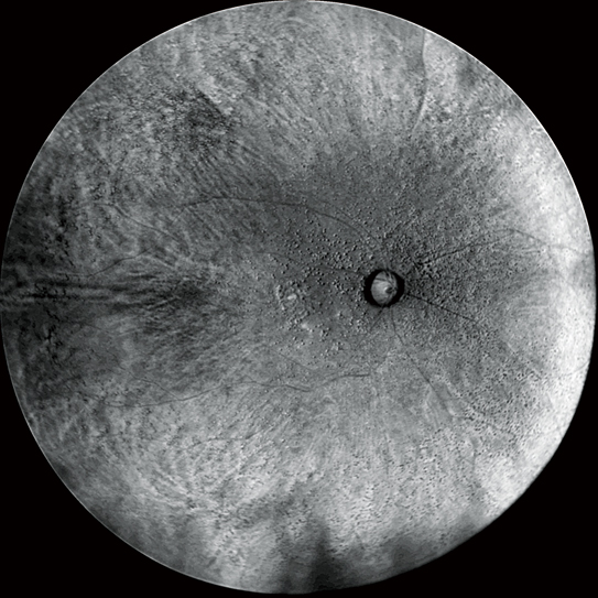
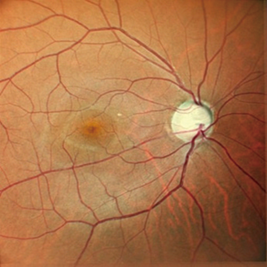
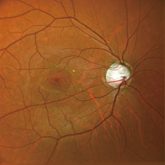
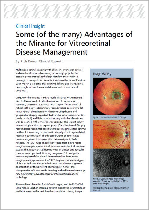
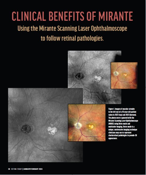
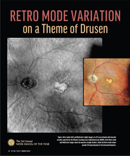
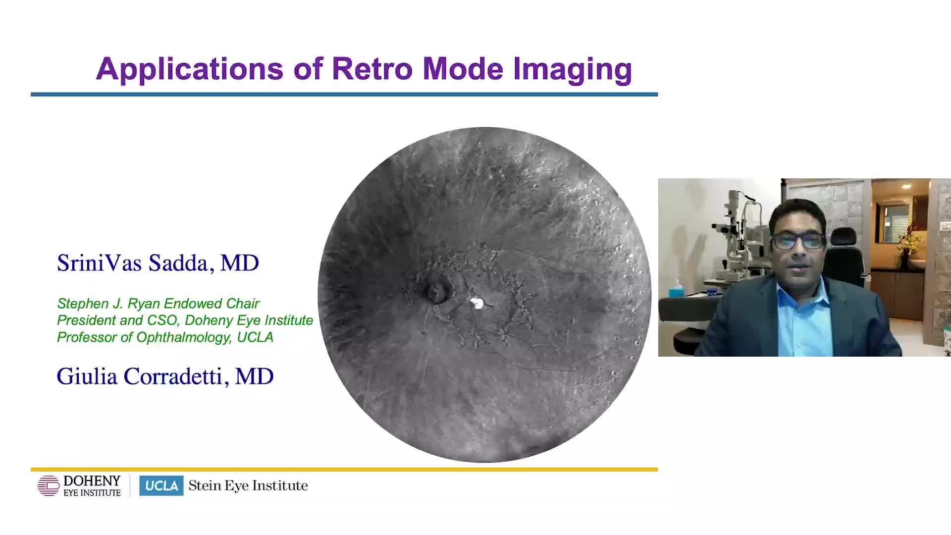
Retro mode
| Retro mode is a unique non-invasive technique for detecting pathologic changes in the choroid. This imaging modality uses scattered IR light to detect abnormal reflection in the choroid caused by drusen, edema and other subtle chorioretinal pathologies. |
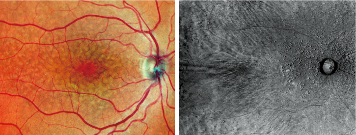
Color and Retro mode images (Drusen) |
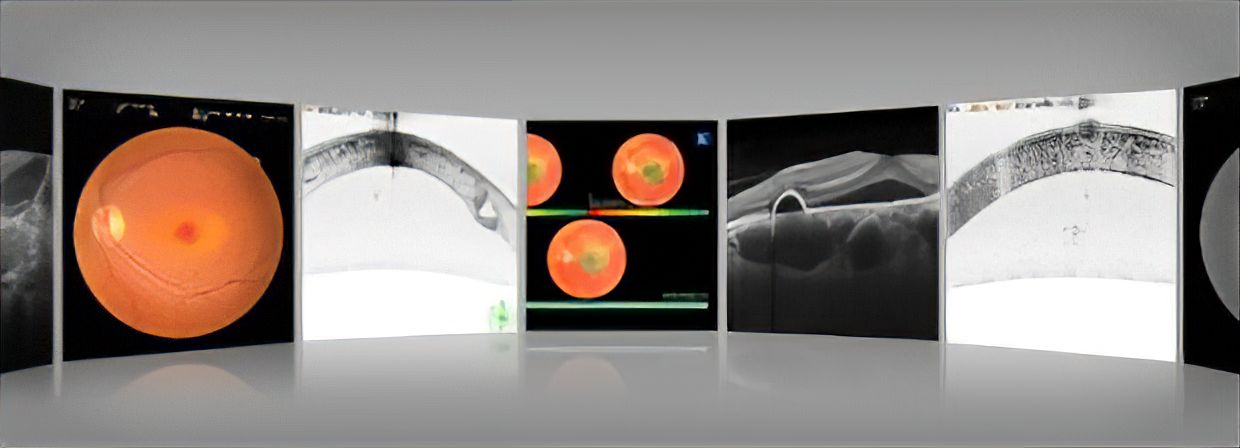
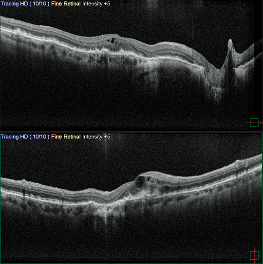
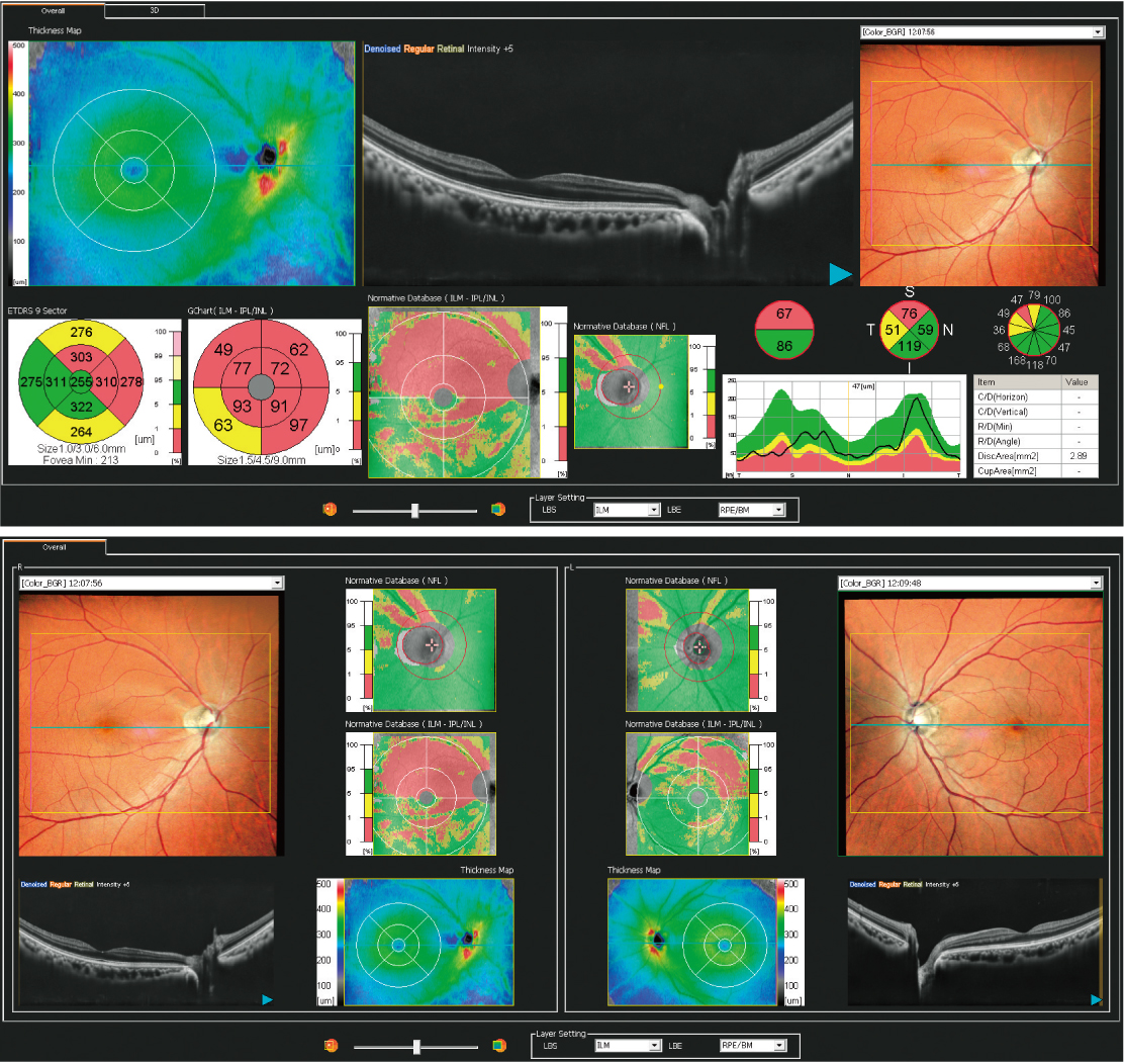
HD wide area OCT*
The Retina map allows wide area diagnosis including the macula and optic disc in a single shot. The ultra fine mode and tracing HD plus functions provide high quality images for detailed observation from vitreous to choroid.
*Available for the SLO/OCT model.
Retina map 12 × 9 mm / 1,024 A-scans x 128 lines |
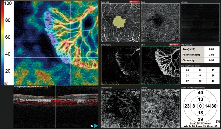
AngioScan OCT-Angiography (optional) |
Fly Through function
| The Fly Through function further enhances multimodal imaging by registering and synchronizing images from different modalities to view the same area while scrolling through the region of interest. | 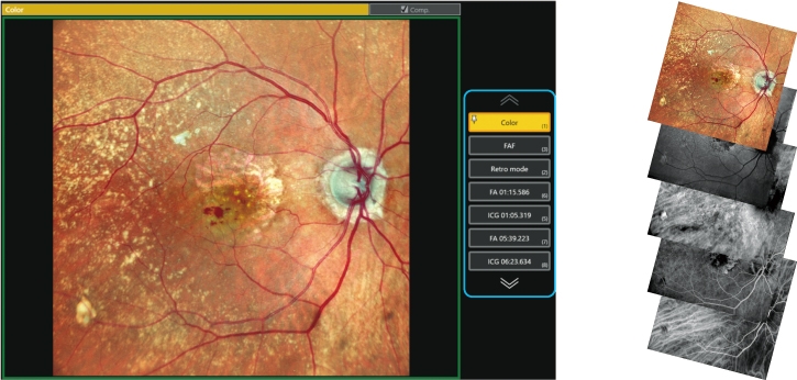 |
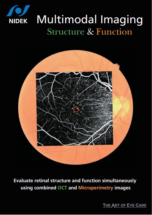
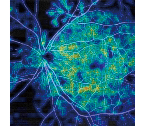
Structural and functional evaluation using Microperimeter*
Various OCT modalities can be registered with microperimetry captured by NIDEK MP-3.
*Available for the SLO/OCT model.
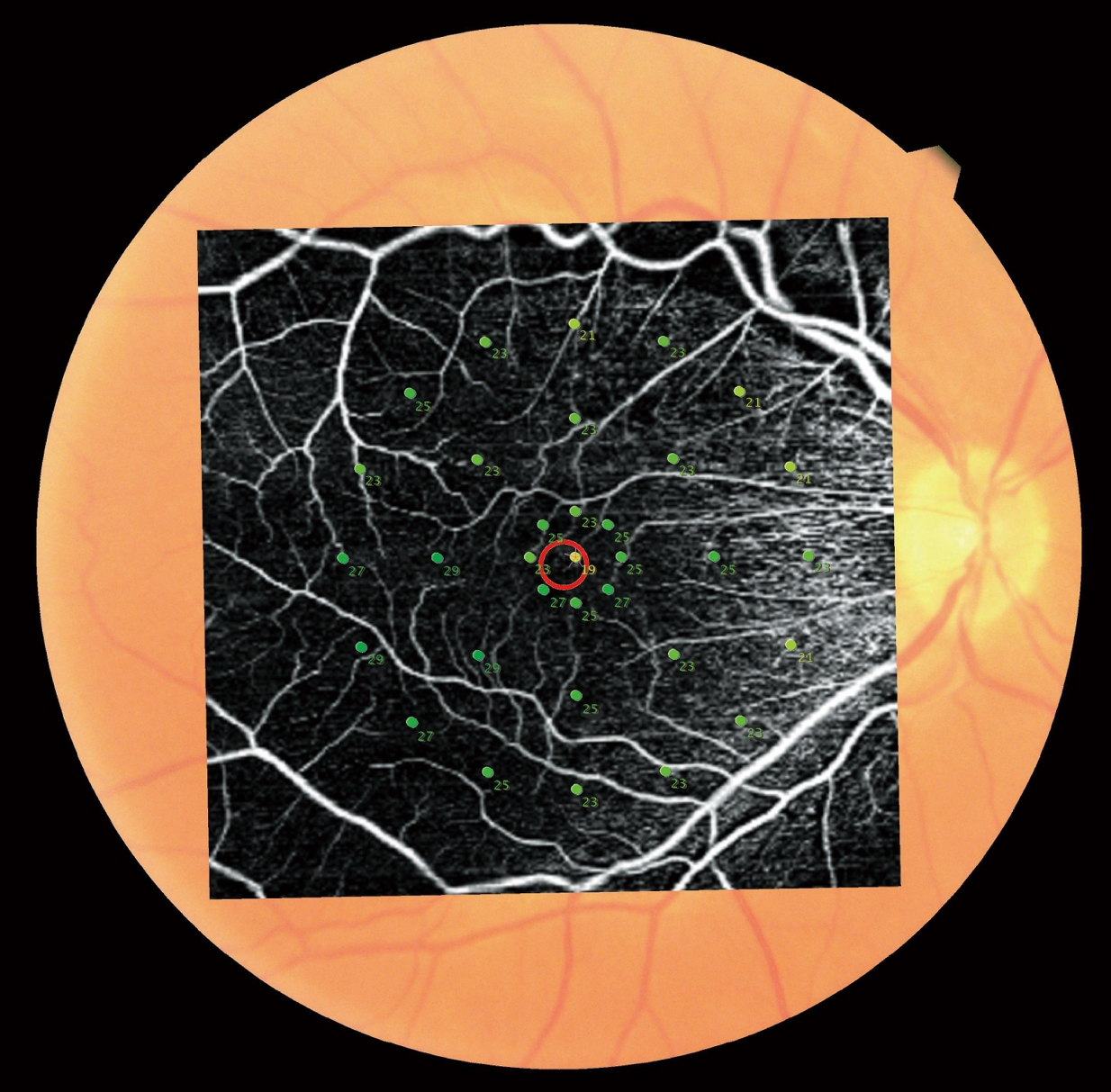
OCT Angiography+Microperimetry |

Normative DB+Microperimetry |
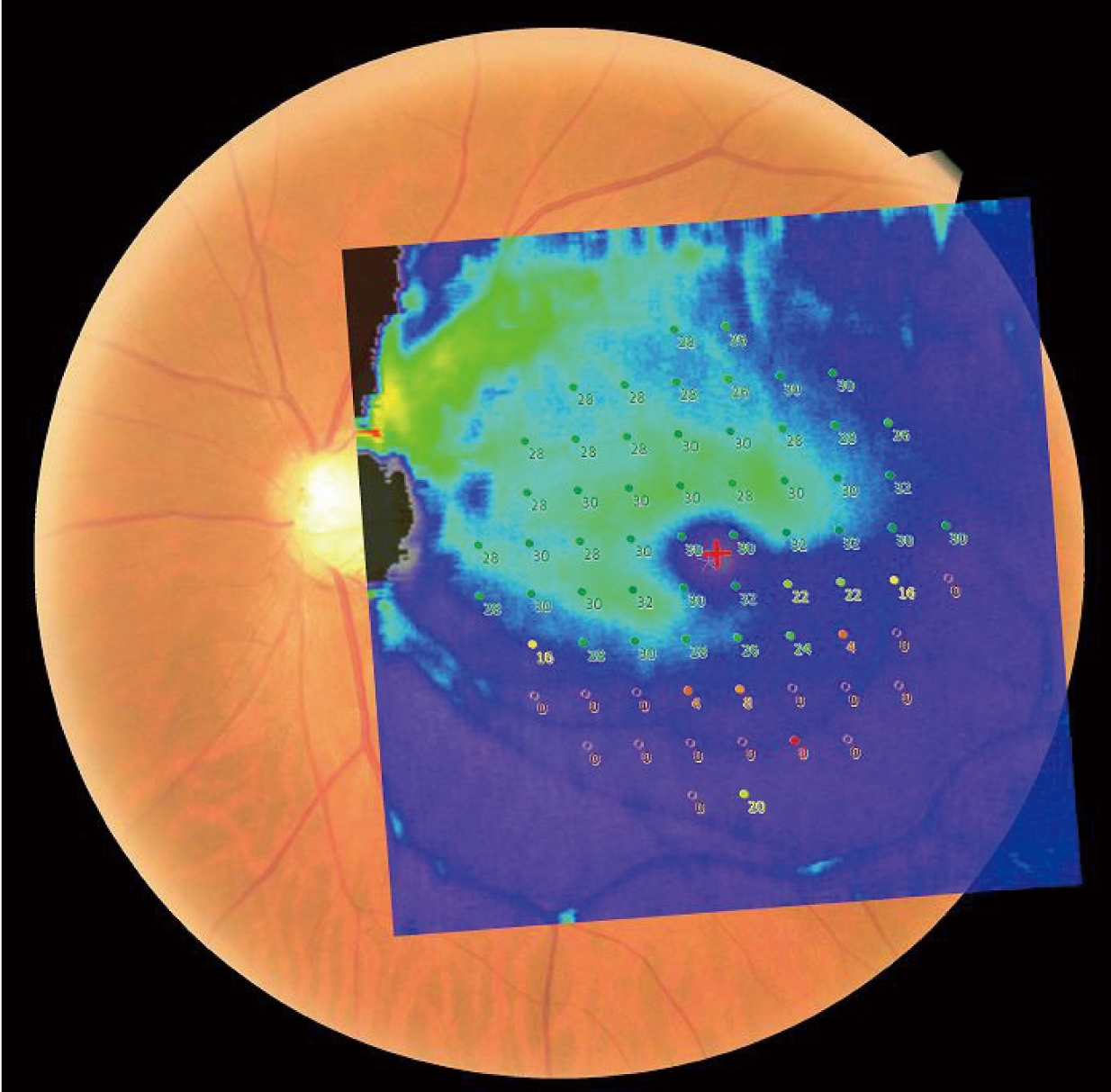
Thickness Map DB+Microperimetry |
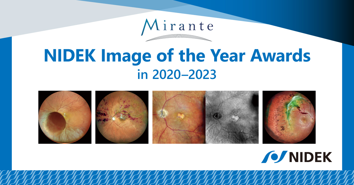
Images courtesy
Luigi Sacco Hospital, University of Milan, Italy
Doheny Eye Center, UCLA, USA
Retina Foundation & Eye Research Center, India
Exilaser Clinic, Peru
Downloads

Multimodal Imaging:Implications for Clinical Practice
|
|
Videos
Product Information
|
Introducción del producto (Spanish)
|
Mirante Viewer / NAVIS-EX Demonstration
|
|
|||
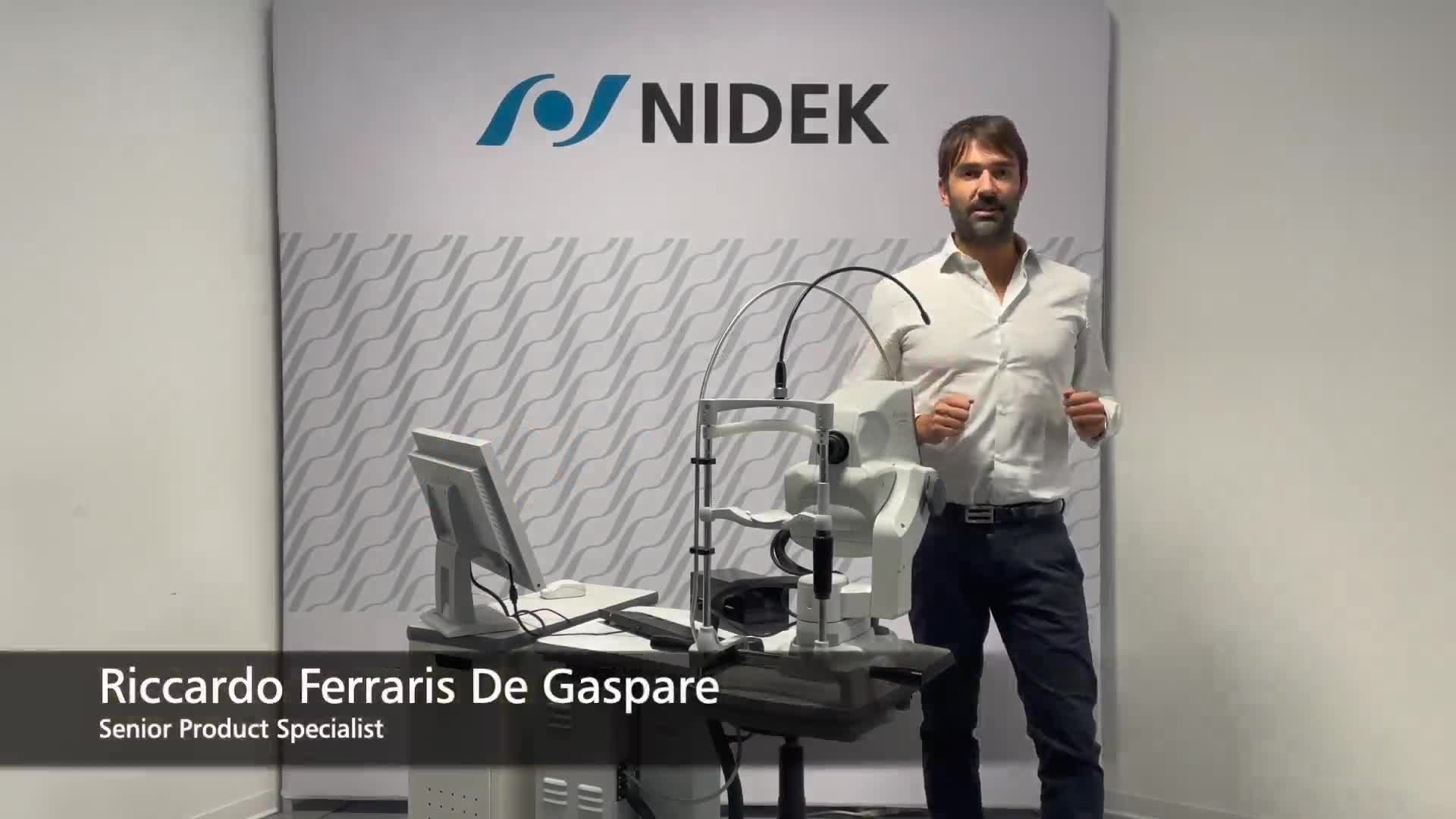
Message from Senior Product Specialist
|
|
Lectures

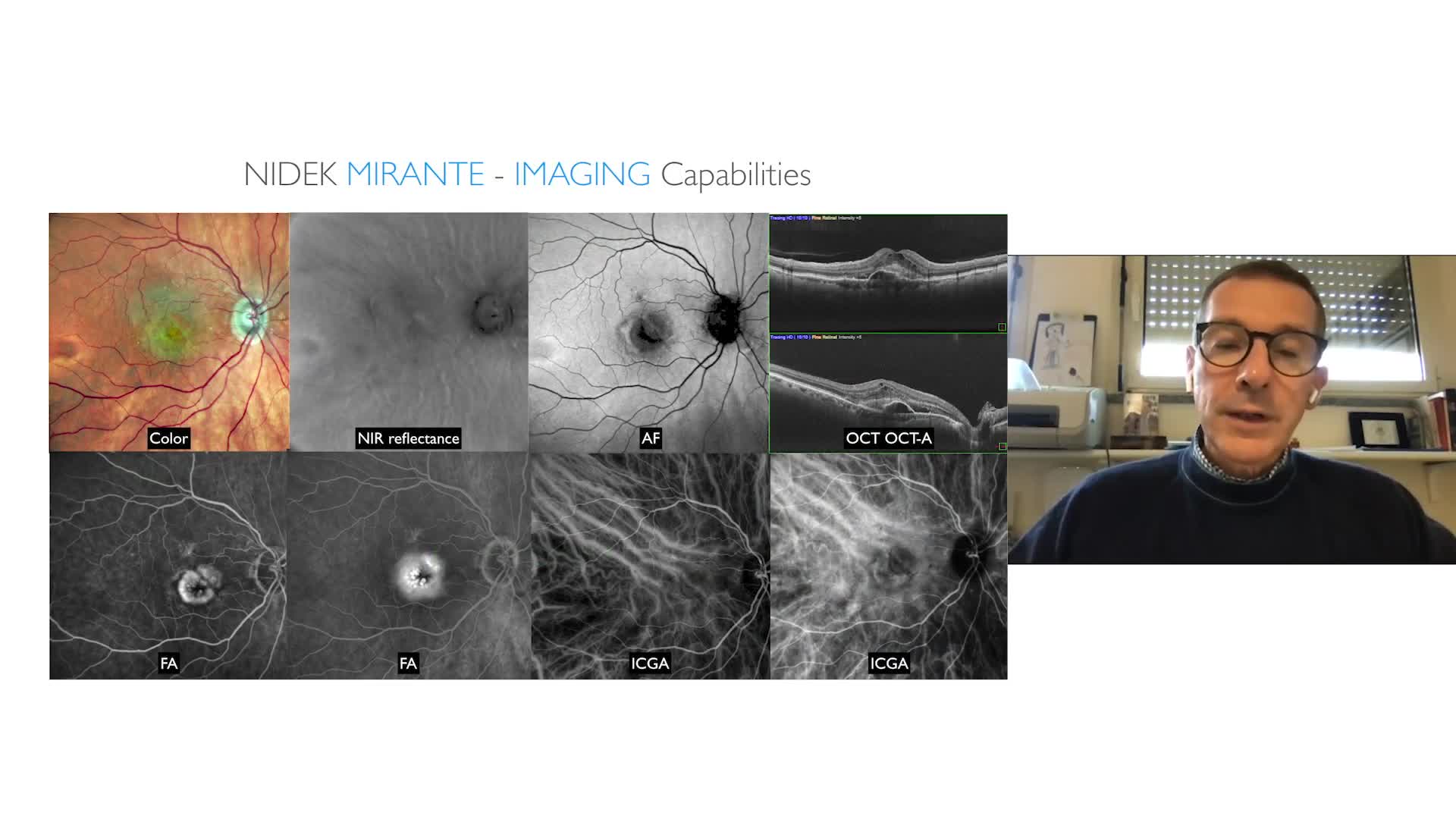
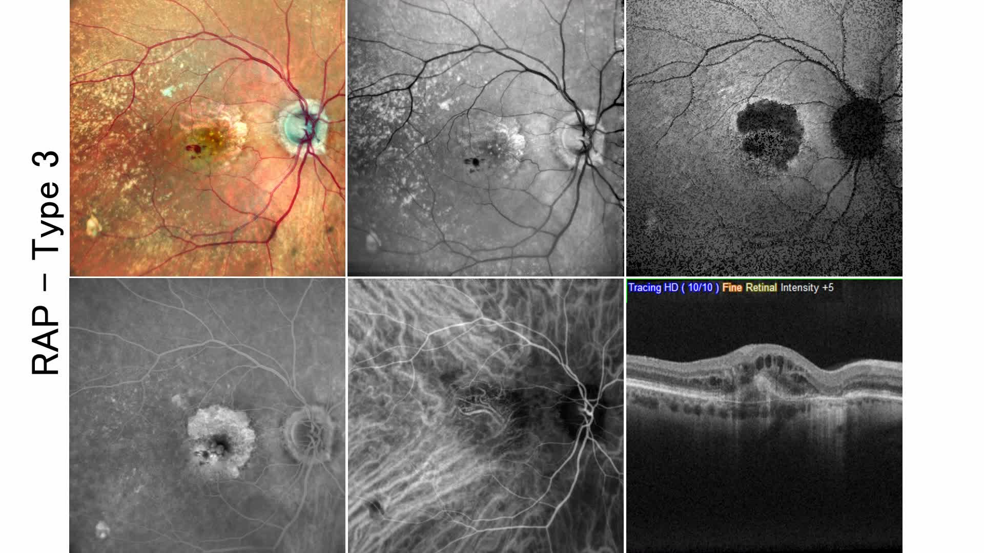
|
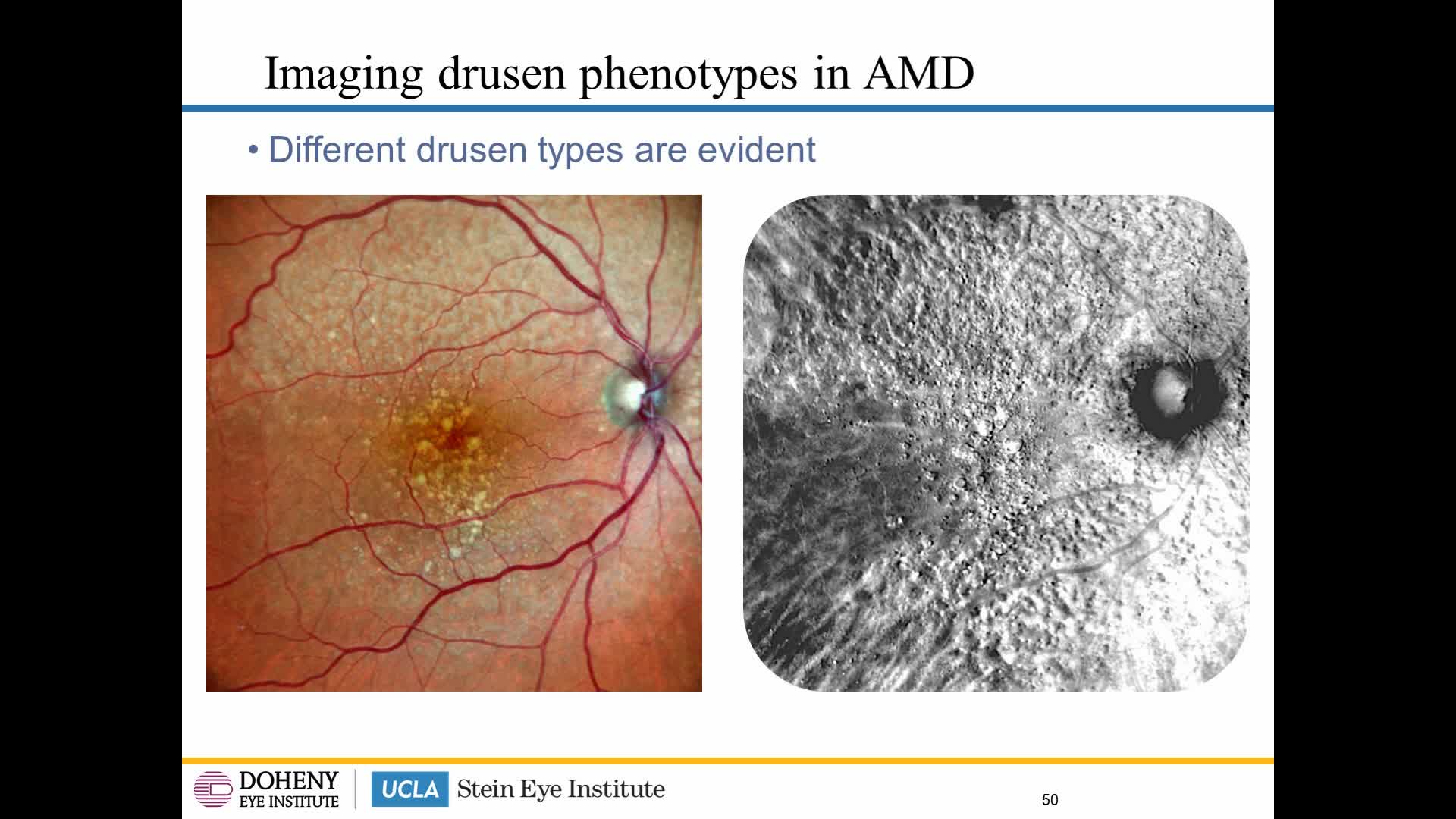
|
|
User Testimonials

Stanislao Rizzo, MD
Professor, Head of Unit of Ophthalmology
Fondazione Policlinico A Gemelli IRCCS & Università Cattolica Sacro Cuore, Italy

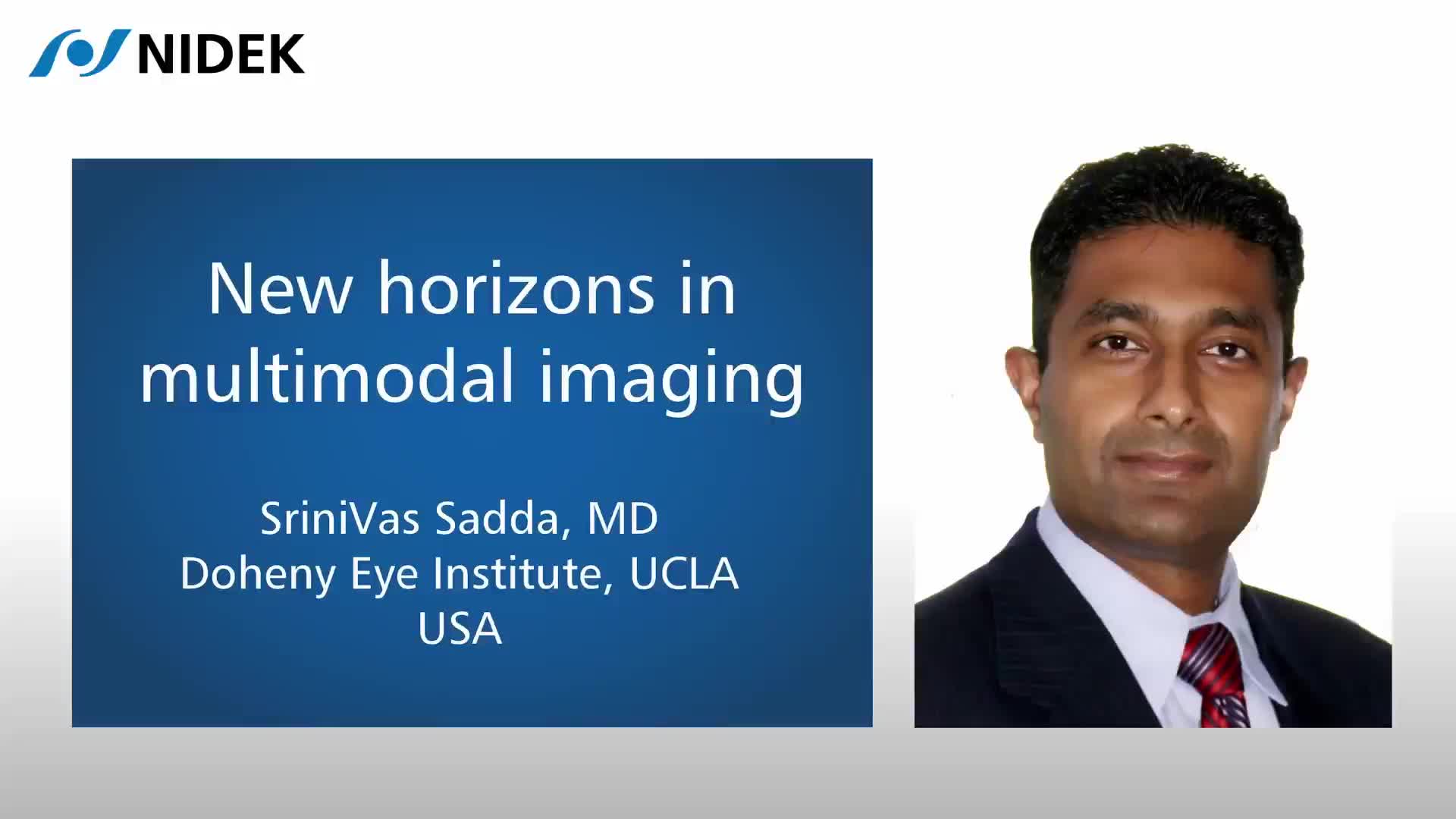
SriniVas R. Sadda, MD
Director, Artificial Intelligence & Imaging Research, Doheny Eye Institute
Professor of Ophthalmology, The University of California – Los Angeles (UCLA), USA

Akihiro Ishibazawa, MD, PhD
Manager of Department of Ophthalmology, Japan Red Cross Kitami Hospital, Japan

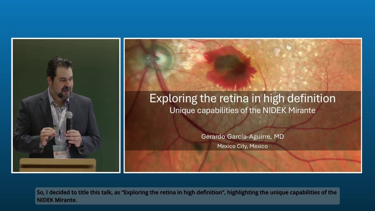
Gerardo Garcia-Aguirre, MD
Attending physician, Asociacion para Evitar la Ceguera en Mexico
Clinical professor of Ophthalmology, School of Medicine and Health Sciences, Tecnologico de Monterrey, Mexico

Manish Nagpal, MD
Senior VR Consultant, Retina Foundation, India

Fabio Patelli, MD
Director, VitreoRetinal service, ASST Santi Paolo e Carlo Hospital, University of Milan
VR consultant, Advalia Vision, Italy

Shozo Sonoda, MD, PhD
Director, Kagoshima Sonoda Eye Clinic & Plastic Surgery
Visiting Professor, Department of Ophthalmology, Kagoshima University, Japan

Nikolle Tan, MD
Consultant Eye Surgeon, Asia Eye Centre, Singapore
Stanislao Rizzo, MD
Professor, Head of Unit of Ophthalmology
Fondazione Policlinico A Gemelli IRCCS & Università Cattolica Sacro Cuore, Italy
SriniVas R. Sadda, MD
Director, Artificial Intelligence & Imaging Research, Doheny Eye Institute
Professor of Ophthalmology, The University of California – Los Angeles (UCLA), USA
Akihiro Ishibazawa, MD, PhD
Manager of Department of Ophthalmology, Japan Red Cross Kitami Hospital, Japan
Gerardo Garcia-Aguirre, MD
Attending physician, Asociacion para Evitar la Ceguera en Mexico
Clinical professor of Ophthalmology, School of Medicine and Health Sciences, Tecnologico de Monterrey, Mexico
Manish Nagpal, MD
Senior VR Consultant, Retina Foundation, India
Fabio Patelli, MD
Director, VitreoRetinal service, ASST Santi Paolo e Carlo Hospital, University of Milan
VR consultant, Advalia Vision, Italy
Shozo Sonoda, MD, PhD
Director, Kagoshima Sonoda Eye Clinic & Plastic Surgery
Visiting Professor, Department of Ophthalmology, Kagoshima University, Japan
Nikolle Tan, MD
Consultant Eye Surgeon, Asia Eye Centre, Singapore
Related Products
OCT-Angiography option for the Mirante SLO/OCT and Retina Scan Duo™ 2
Optical Coherence Tomography
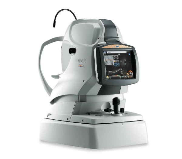
RS-1 Glauvas
Optical Coherence Tomography / Fundus Camera
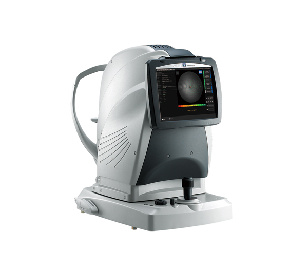
Retina Scan Duo™2
Software for NIDEK OCT series
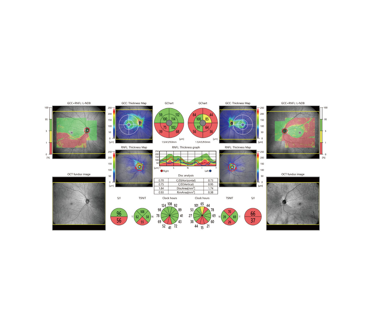
Long Axial Length Normative Database
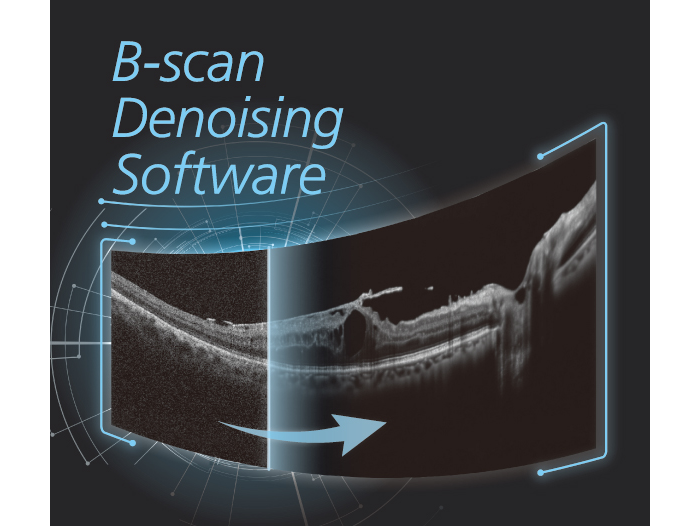
B-scan Denoising Software
for NIDEK OCT series
Microperimeter
MP-3
Non-mydriatic Auto Fundus Camera
AFC-330
NOTE
The availability of products differs from country to country depending on the status of approval.
Specifications and design are subject to change without notice.
- Product/model name
- Scanning Laser Ophthalmoscope Mirante